. 2017 Oct 23;12(10):e0186511.
doi: 10.1371/journal.pone.0186511. eCollection 2017.

Protective effect of dexamethasone on 5-FU-induced oral mucositis in hamsters

Affiliations

Protective effect of dexamethasone on 5-FU-induced oral mucositis in hamsters

Susana Barbosa Ribeiro et al. PLoS One. .

Abstract

Oral mucositis (OM) is an important side effect of cancer treatment, characterized by ulcerative lesions in the mucosa of patients undergoing radiotherapy or chemotherapy, which has marked effects on patient quality of life and cancer therapy continuity. Considering that few protocols have demonstrated efficacy in preventing this side effect, the aim of this study was to examine the effect of dexamethasone (DEX) on OM induced by 5-fluorouracil (5-FU) in hamsters by studying signaling pathways. OM was induced in hamsters by 5-FU followed by mechanical trauma (MT) on day 4. On day 10, the animals were euthanized. The experimental groups included saline, MT, 5-FU, and DEX (0.25, 0.5, or 1 mg/kg). Macroscopic, histopathological, and immunohistochemical analyses as well as immunofluorescence experiments were performed on the oral mucosa of the animals. The oral mucosal samples were analyzed by enzyme-linked immunosorbent assays, and quantitative real-time polymerase chain reaction (qPCR). DEX (0.5 or 1 mg/kg) reduced inflammation and ulceration of the oral mucosa of hamsters. In addition, DEX (1 mg/kg) reduced the cytokine levels of tumor necrosis factor (TNF)-α, interleukin (IL)-1β, and macrophage migration inhibitory factor (MIF). DEX (1 mg/kg) also reduced the immunoexpression of cyclooxygenase (COX)-2, matrix metalloproteinase (MMP)-2, transforming growth factor (TGF)-β, MIF, Smad 2/3, Smad 2/3 phosphorylated and NFκB p65 in the jugal mucosa. Finally, DEX (1 mg/kg) increased interleukin-1 receptor-associated kinase 3 (IRAK-M), glucocorticoid-induced leucine zipper (GILZ), and mitogen-activated protein kinase (MKP1) gene expression and reduced NFκB p65 and serine threonine kinase (AKt) gene expression, relative to the 5-FU group. Thus, DEX improved OM induced by 5-FU in hamsters.


Conflict of interest statement

Competing Interests: The authors have declared that no competing interests exist.

Figures

Fig 1
Fig 1
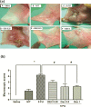
Dexamethasone (DEX) improved the macroscopic analysis (a) and scores (b) of the oral mucosa of hamsters with oral mucositis (OM) induced by 5-fluorouracil (5-FU) and MT. The saline group consisted of normal animals without OM induction (A). The mechanical trauma (MT) group received excoriations in the jugal mucosa, without 5-FU treatment (B). The 5-FU group received 5-FU, was subjected to MT, and was treated with saline i.p. (C). The DEX groups received 5-FU, were subjected to MT, and received DEX i.p. at one of the three different doses 0.25 (D), 0.5 (E), or 1 mg/kg (F). Scores are represented as the median ± standard error of the mean (n = 5). *p < 0.05 vs. the saline group, #p < 0.05 vs. the 5-FU group (Kruskal–Wallis test and Dunn’s multiple comparison test).
Fig 2
Fig 2
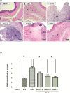
Histopathological analysis (a) and scores (b) of the oral mucosa of hamsters with oral mucositis (OM) induced by 5-fluorouracil (5-FU). The saline group consisted of normal animals without OM and without mucosal alterations (A). The mechanical trauma (MT) group received excoriations in the jugal mucosa, without 5-FU treatment (B). The 5-FU group received 5-FU, was subjected to MT, and was treated with saline i.p.; this group had changes in the jugal mucosa (C), with the presence of ulcers (star) and inflammatory infiltration (two asterisks). The dexamethasone (DEX) groups received 5-FU, were subjected to MT, and received DEX i.p. at one of three different doses: 0.25 mg/kg (D), 0.5 mg/kg (E), or 1 mg/kg (F) with mild hyperemia (an arrow) and absence of ulcers. Scores are represented as the median ± standard error of the mean (n = 5). *p < 0.05 vs. the saline group, #p < 0.05 vs. the 5-FU group (Kruskal–Wallis test and Dunn’s multiple comparison test).
Fig 3
Fig 3
Tumor necrosis factor (TNF)-α (a) and interleukin (IL)-1β (b) cytokines in the oral mucosa of hamsters with oral mucositis (OM). The saline group consisted of normal animals without OM. The mechanical trauma (MT) group consisted of hamsters receiving excoriations in the oral mucosa, without 5-fluorouracil (5-FU) treatment. The 5-FU group received 5-FU, was subjected to MT, and was treated with saline i.p. The dexamethasone (DEX) groups received 5-FU, were subjected to MT, and received DEX i.p. at one of three different doses (0.25, 0.5, or 1 mg/kg). The results are presented as the mean ± standard error of the mean (n = 5). *p < 0.05 vs. the saline group, #p < 0.05 vs. the 5-FU group (analysis of variance with Tukey’s post-test).
Fig 4
Fig 4
Immunohistochemistry (a) and scores (b) for matrix metalloproteinase-2 (MMP-2), cyclooxygenase-2 (COX-2), and transforming growth factor beta (TGF-β). The saline group (A, E, and I) and the mechanical trauma (MT) group (B, F, and J) had little immunostaining. The 5-fluorouracil (5-FU) group had intense labeling in the jugal mucosa for MMP-2 (C), COX-2 (G), and TGF-β (K), compared to the saline group. Dexamethasone (DEX, 1 mg/kg) reduced the immunostaining for MMP-2 (D), COX-2 (H), and TGF-β (L), compared to the 5-FU group. Poor immunoblotting (an asterisk) and intense immunostaining (two asterisks). Scores are represented as the median ± standard error of the mean (n = 5). *p < 0.05 vs. the saline group, #p < 0.05 vs. the 5-FU group (Kruskal-Wallis test and Dunn’s multiple comparison test).
Fig 5
Fig 5
Immunofluorescence for MIF and NFκB p65 (a) and mean of densitometric analysis (b). The 5-fluorouracil (5-FU) group (C and G) had higher green labeling than in the mechanical trauma (MT) group (B and F) or the saline group (A and E) (p <0.05; n = 5). DEX 1 mg/kg group (D and H) had a reduced immunoreactivity as compared to the 5-FU group (n = 5; *p < 0.05 vs. the saline group, #p < 0.05 vs. the 5-FU group; (analysis of variance with Tukey’s post-test).
Fig 6
Fig 6
Immunofluorescence for pSmad 2/3 and Smad2/3 (a) and mean of densitometric analysis (b). The 5-fluorouracil (5-FU) group (C and G) had higher green labeling than in the mechanical trauma (MT) group (B and F) or the saline group (A and E) (p <0.05; n = 5). DEX 1 mg/kg group (D and H) had a reduced immunoreactivity as compared to the 5-FU group (n = 5; *p < 0.05 vs. the saline group, #p < 0.05 vs. the 5-FU group; (analysis of variance with Tukey’s post-test).
Fig 7
Fig 7
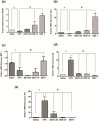
Real-time polymerase chain reaction for interleukin-1 receptor-associated kinase 3 (IRAK-M) (a), glucocorticoid-induced leucine zipper (GILZ) (b), MAPK phosphatase 1 (MKP1) (c), nuclear factor NFκB p65 (d), and serine threonine kinase (AKt) (e). 5-Fluorouracil (5-FU) decreased IRAK-M, GILZ, and MKP1 gene expression as well as increased NFκB p65 and AKt gene expression, compared to the saline group. Dexamethasone (DEX, 1 mg/kg) increased IRAK-M, GILZ, and MKP1 gene expression as well as reduced NFκB p65 and AKt gene expression, compared to the 5-FU group (n = 5; *p < 0.05 vs. the saline group, #p < 0.05 vs. the 5-FU group; analysis of variance with Tukey’s post-test).
Fig 8
Fig 8. Signaling pathways of dexamethasone modulation in oral mucositis induced by 5-fluorouracil (5-FU).
5-FU activates NFkB which induces expression of pro-inflammatory cytokines (IL-1 β, TNF-α) that promote tissue damage and inflammation in the oral mucosa. Dexamethasone downregulated the NFkB by increasing the expression of IRAKM and GILZ and decreased gene expression of AKt. Dexamethasone interfering with NFkB decreases IL-1 β, TNF-α, TGF-β, COX2 and MMP2 by improving oral mucositis. (NFκB—the nuclear transcription factor kappa B; GILZ- glucocorticoid-induced leucine zipper; IRAK-M—interleukin-1 receptor-associated kinase; AKt- serine threonine kinase).

References

    1. Silverman S Jr. (2007) Diagnosis and management of oral mucositis. J Support Oncol 5: 13–21. - PubMed
    1. Lalla RV, Sonis ST, Peterson DE (2008) Management of oral mucositis in patients who have cancer. Dent Clin North Am 52: 61–77, viii. doi: 10.1016/j.cden.2007.10.002 - DOI - PMC - PubMed
    1. Sonis ST (2010) New thoughts on the initiation of mucositis. Oral Dis 16: 597–600. doi: 10.1111/j.1601-0825.2010.01681.x - DOI - PubMed
    1. Sonis ST (2007) Pathobiology of oral mucositis: novel insights and opportunities. J Support Oncol 5: 3–11. - PubMed
    1. Logan RM, Gibson RJ, Sonis ST, Keefe DM (2007) Nuclear factor-kappaB (NF-kappaB) and cyclooxygenase-2 (COX-2) expression in the oral mucosa following cancer chemotherapy. Oral Oncol 43: 395–401. doi: 10.1016/j.oraloncology.2006.04.011 - DOI - PubMed