

Central sulcus development in early childhood
- PMID: 29059835
- PMCID: PMC6554210
- DOI: 10.1109/EMBC.2017.8036787
Central sulcus development in early childhood
Abstract
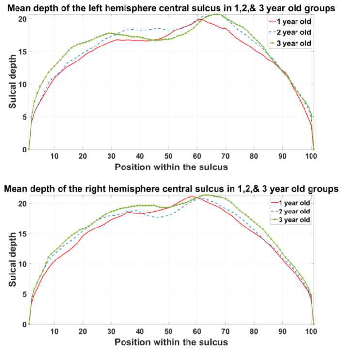
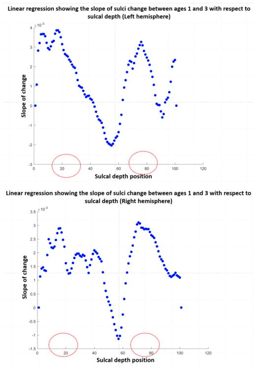
Mapping out the development of the brain in early childhood is a critical part of understanding neurological disorders. The brain grows rapidly in early life, reaching 95% of the final volume by age 6. A normative atlas containing structural parameters that indicate development would be a powerful tool in understanding the progression of neurological diseases. Although some studies have begun exploring cortical development in pediatric imaging, sulci have not been examined extensively. Here, we study the changes in the Central Sulcus (CS), which is one of the earliest sulci to develop from the fetal stage, at early developmental age 1-3 years old using high resolution magnetic resonance images. Parameterization of the central sulcus was performed and results show us that the CS change corresponds to the development of the mouth and tongue regions.
Figures

References
-
- Leroy F, Cai Q, Bogart SL, Dubois J, Coulon O, Monzalvo K, Fischer C, Glasel H, Van der Haegen L, Bénézit A, Lin C-P, Kennedy DN, Ihara AS, Hertz-Pannier L, Moutard M-L, Poupon C, Brysbaert M, Roberts N, Hopkins WD, Mangin J-F, Dehaene-Lambertz G. New human-specific brain landmark: the depth asymmetry of superior temporal sulcus. Proc Natl Acad Sci U S A. 2015 Jan;112(4):1208–13. - PMC - PubMed
-
- Coulon O, Lefevre J, Kloppel S, Siebner H, Mangin J-F. Quasi-isometric length parameterization of cortical sulci: Application to handedness and the central sulcus morphology. 2015 IEEE 12th International Symposium on Biomedical Imaging (ISBI); 2015; pp. 1268–1271.
-
- Sun ZY, Klöppel S, Rivière D, Perrot M, Frackowiak R, Siebner H, Mangin J-F. The effect of handedness on the shape of the central sulcus. Neuroimage. 2012 Mar;60(1):332–9. - PubMed
MeSH terms
Grants and funding
LinkOut - more resources
Full Text Sources
Other Literature Sources