

doi: 10.1109/EMBC.2017.8037113.
Multi-scale locally low-rank noise reduction for high-resolution dynamic quantitative cardiac MRI
- PMID: 29060157
- PMCID: PMC6938225
- DOI: 10.1109/EMBC.2017.8037113
Item in Clipboard
Multi-scale locally low-rank noise reduction for high-resolution dynamic quantitative cardiac MRI
Annu Int Conf IEEE Eng Med Biol Soc.
2017 Jul.
Abstract
Evaluation of myocardial T1 times is conventionally limited to a single temporal snapshot of the cardiac cycle, leaving the dependence between functional and tissue characterization unexplored. We recently proposed a technique that alleviates this limitation by acquiring dynamic quantitative myocardial T1 maps. However, tradeoffs between temporal resolution, scan duration and SNR limit the spatial resolution. In this work, we propose a multi-scale locally low rank noise reduction approach without parameter-tuning to enable high acceleration rates in the acquisition, facilitating superior spatial and temporal resolutions in dynamic myocardial T1 mapping.
Figures
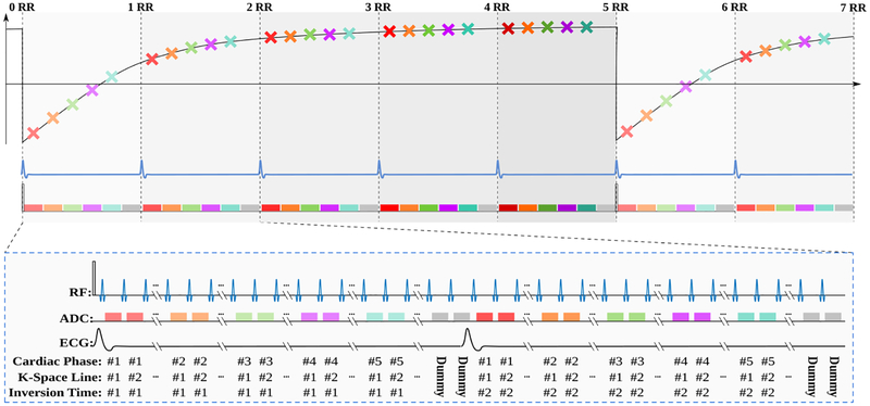
The sequence diagram of the dynamic myocardial T1 mapping used in this study. Overall, the sequence acquires ncardiac cardiac phases per heart beat (five in the figure, depicted with different colors) over ncontrast heartbeats (five in the figure) that is necessary until re-recovery to pulsed steady-state for a FLASH sequence. This results in ncardiac
· ncontrast images in a series that varies both in contrast and cardiac motion.

Results of denoising on baseline with in-plane resolution of 1.3 × 1.3 mm2, reconstructed with GRAPPA (unprocessed) and with the proposed noise variance reduction technique (proposed).

Dynamic quantitative T1 maps generated without and with the proposed multi-scale LLR noise reduction technique. Homogeneous T1 values are observed with the proposed approach, with > 60% reduction in spatial variability.

T1 times through cardiac phases across a cross-section of the heart, showing no temporal blurring occurs with the proposed technique.
References
-
- von Knobelsdorff-Brenkenhoff F and Schulz-Menger J, “Cardiovascular magnetic resonance imaging in ischemic heart disease,” J Magn Reson Imaging, 36(1):20–38, 2012. - PubMed
-
- Schelbert EB and Messroghli DR DR, “State of the Art: Clinical Applications of Cardiac T1 Mapping,” Radiology, 278:658–676, 2016. - PubMed
-
- Messroghli DR, Radjenovic A, Kozerke S et al., “Modified Look-Locker inversion recovery (MOLLI) for high-resolution T1 mapping of the heart,” Magn Reson Med, 52(1):141–146, 2004. - PubMed
-
- Chow K, Flewitt JA, Green JD et al., “Saturation recovery single-shot acquisition (SASHA) for myocardial T1 mapping,” Magn Reson Med, 71(6):2082–2095, 2014. - PubMed
MeSH terms
Grants and funding
LinkOut - more resources
Full Text Sources
Other Literature Sources