. 2017 Oct 23;14(1):205.
doi: 10.1186/s12974-017-0977-4.

Elevated microRNA-129-5p level ameliorates neuroinflammation and blood-spinal cord barrier damage after ischemia-reperfusion by inhibiting HMGB1 and the TLR3-cytokine pathway

Affiliations

Elevated microRNA-129-5p level ameliorates neuroinflammation and blood-spinal cord barrier damage after ischemia-reperfusion by inhibiting HMGB1 and the TLR3-cytokine pathway

Xiao-Qian Li et al. J Neuroinflammation. .

Erratum in

Abstract

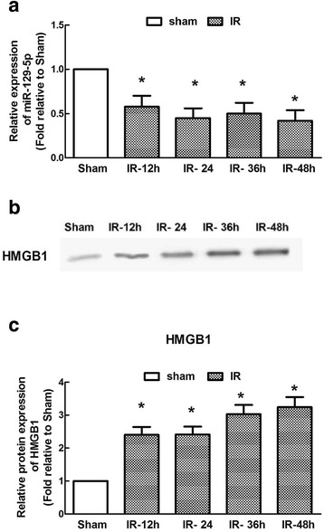
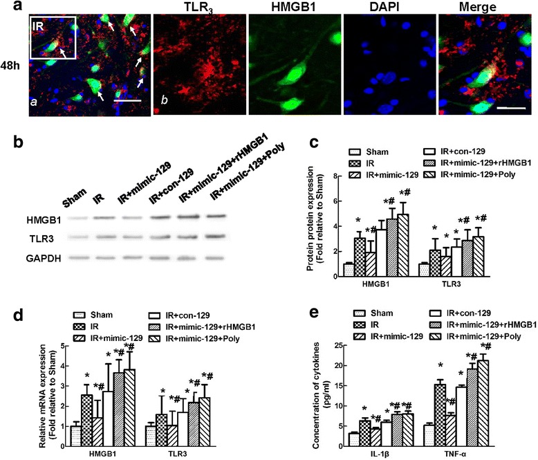
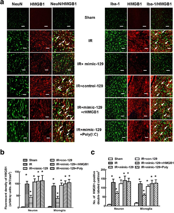
Background: Ischemia-reperfusion (IR) affects microRNA (miR) expression and causes substantial inflammation. Multiple roles of the tumor suppressor miR-129-5p in cerebral IR have recently been reported, but its functions in the spinal cord are unclear. Here, we investigated the role of miR-129-5p after spinal cord IR, particularly in regulating high-mobility group box-1 (HMGB1) and the Toll-like receptor (TLR)-3 pathway.

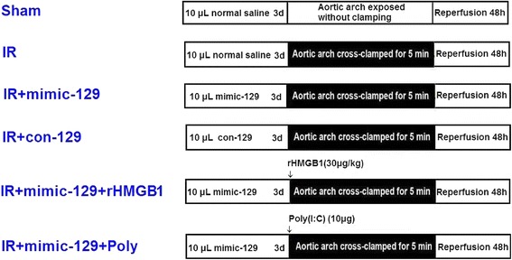
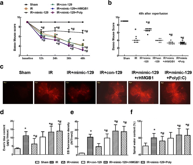
Methods: Ischemia was induced via 5-min occlusion of the aortic arch. The relationship between miR-129-5p and HMGB1 was elucidated via RT-PCR, western blotting, and luciferase assays. The cellular distribution of HMGB1 was determined via double immunofluorescence. The effect of miR-129-5p on the expression of HMGB1, TLR3, and downstream cytokines was evaluated using synthetic miRs, rHMGB1, and the TLR3 agonist Poly(I:C). Blood-spinal cord barrier (BSCB) permeability was examined by measuring Evans blue (EB) dye extravasation and the water content.

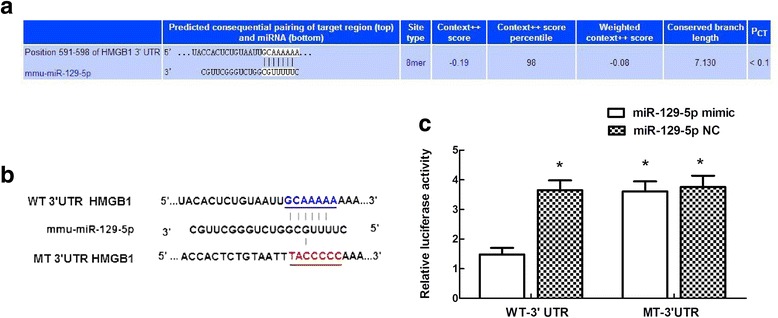
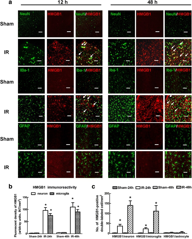
Results: The temporal miR-129-5p and HMGB1 expression profiles and luciferase assay results indicated that miR-129-5p targeted HMGB1. Compared with the Sham group, the IR group had higher HMGB1 immunoreactivity, which was primarily distributed in neurons and microglia. Intrathecal injection of the miR-129-5p mimic significantly decreased the HMGB1, TLR3, interleukin (IL)-1β and tumor necrosis factor (TNF)-α levels and the double-labeled cell count 48 h post-surgery, whereas rHMGB1 and Poly(I:C) reversed these effects. Injection of miR-129-5p mimic preserved motor function and prevented BSCB leakage based on increased Basso Mouse Scale scores and decreased EB extravasation and water content, whereas injection rHMGB1 and Poly(I:C) aggravated these injuries.

Conclusions: Increasing miR-129-5p levels protect against IR by ameliorating inflammation-induced neuronal and BCSB damage by inhibiting HMGB1 and TLR3-associated cytokines.

Keywords: Blood-spinal cord barrier; High-mobility group box-1; Ischemia-reperfusion injury; MicroRNAs; Toll-like receptor 3.


Conflict of interest statement

Ethics approval

All animal experiments were approved by the Ethics Committee of China Medical University and performed in compliance with institutional guidelines under approved protocols.

Consent for publication

Not applicable

Competing interests

The authors declare that they have no competing interests.

Publisher’s Note

Springer Nature remains neutral with regard to jurisdictional claims in published maps and institutional affiliations.

Figures

Fig. 1
Fig. 1
Experimental groups and protocol. Schematic representation of the six groups of mice exposed to the different treatments
Fig. 2
Fig. 2
Time course of IR-induced alterations in the miR-129-5p and HMGB1 levels in a mouse model of spinal cord IR injury. a Quantification of miR-129-5p expression. b Representative western blots to detect HMGB1 expression. c Quantification of the integrated intensity of the HMGB1 bands. Data are expressed as the mean ± SD. n = 6 per group. *P < 0.05 versus the Sham group
Fig. 3
Fig. 3
Confirmation of a direct interaction between miR-129-5p and HMGB1. a Schematic representation of the predicted interaction between miR-129-5p and HMGB1 based on TargetScan. b Binding site of miR-129-5p in the WT and MT 3′UTR of HMGB1. c Dual-luciferase reporter gene assay. Luciferase activity was measured after 48 h of co-transfection of HEK-293 cells with the miR-129-5p mimic and the WT or MT reporter vector. Compared to the control, miR-129-5p significantly decreased the luciferase activity of the WT vector but not that of the MT vector. Data are expressed as the mean ± SD. *P < 0.05 versus the control miR
Fig. 4
Fig. 4
Double immunofluorescence of HMGB1 with markers of major cell types in the spinal cord after IR. a Representative fluorescence images of the distribution of HMGB1 (red) in neurons (NeuN; green), microglia (Iba1; green) and astrocytes (GFAP; green) in the spinal cords at 12 and 48 h after IR. Arrows indicate co-localization. Scale bars = 50 μm. b Quantification of HMGB1 signals was performed based on the average of three independent images. c Quantification of HMGB1-positive neurons and microglia in the spinal cords at 12 h and 48 h after IR. Data are expressed as the mean ± SD. *P < 0.05 versus the Sham group
Fig. 5
Fig. 5
Effect of the miR-129-5p mimic and mimic control on the expression of HMGB1 and TLR3-mediated cytokines at 48 h after IR. a Representative fluorescence images of the distribution of TLR3(red), HMGB1(green) and nuclei (blue) in the spinal cords at 48 h after IR. b Representative western blots of HMGB1 and TLR3 expression. GAPDH was used as a loading control. c Quantification of the relative protein expression of HMGB1 and TLR3 at 48 h after IR. d Quantification of the relative mRNA expression of HMGB1 and TLR3 at 48 h after IR. e Quantification of spinal IL-1β and TNF-α production at 48 h after IR. All data were obtained from three independent experiments and expressed as the mean ± SD. *P < 0.05 versus the Sham group. # P < 0.05 versus the IR group
Fig. 6
Fig. 6
Effects of the miR-129-5p mimic and mimic control on HMGB1 expression in specific cell types of the spinal cord after IR. a Representative photomicrographs showing the localization of the fluorescence signals for HMGB1 in neurons and microglia at 48 h after IR. Arrows indicate co-localization. Scale bars = 50 μm. b Quantification of HMGB1 signals was performed based on the average of three independent images. c Quantification of HMGB1-positive neurons and microglia in the spinal cords at 48 h after IR. Data are expressed as the mean ± SD. *P < 0.05 versus the Sham group. # P < 0.05 versus the IR group
Fig. 7
Fig. 7
Effects of the miR-129-5p mimic and mimic control on hind-limb motor function and blood-spinal cord barrier (BSCB) integrity after IR. a Hind-limb motor function was assessed at 12-h intervals for 48 h after reperfusion using the Basso Mouse Scale for locomotion with a range from 0 (paraplegia) to 9 (normal). b Assessment of each mouse at 48 h post-surgery in the six groups. Each symbol represents one mouse. The bar represents the median. c Changes in BSCB permeability were measured based on EB extravasation visualized as red fluorescence by microscopy. d Quantification of the EB content of the spinal cord (μg/g tissue). e Quantification of the EB fluorescence intensity (INT/mm2). f Quantification of the spinal water content (%). Data are expressed as the mean ± SD. *P < 0.05 versus the Sham group. # P < 0.05 versus the IR group

References

    1. He F, Shi E, Yan L, Li J, Jiang X. Inhibition of micro-ribonucleic acid-320 attenuates neurologic injuries after spinal cord ischemia. J Thorac Cardiovasc Surg. 2015;150:398–406. doi: 10.1016/j.jtcvs.2015.03.066. - DOI - PubMed
    1. Li XQ, Lv HW, Tan WF, Wang ZL, Fang B, Ma H. MiR-27a ameliorates inflammatory damage to the blood-spinal cord barrier after spinal cord ischemia-reperfusion injury in rats by down-regulating TICAM-2 of the TLR4 signaling pathway. J Neuroinflammation. 2015;12:25. doi: 10.1186/s12974-015-0246-3. - DOI - PMC - PubMed
    1. Fang B, Li XQ, Bao NR, Tan WF, Chen FS, Pi XL, et al. Role of autophagy in the bimodal stage after spinal cord ischemia reperfusion injury in rats. Neuroscience. 2016;328:107–116. doi: 10.1016/j.neuroscience.2016.04.019. - DOI - PubMed
    1. Bell MT, Puskas F, Agoston VA, Cleveland JC, Jr, Freeman KA, Gamboni F, et al. Toll-like receptor 4-dependent microglial activation mediates spinal cord ischemia-reperfusion injury. Circulation. 2013;128(11 Suppl 1):S152–S156. doi: 10.1161/CIRCULATIONAHA.112.000024. - DOI - PubMed
    1. Li XQ, Wang J, Fang B, Tan WF, Ma H. Intrathecal antagonism of microglial TLR4 reduces inflammatory damage to blood-spinal cord barrier following ischemia/reperfusion injury in rats. Mol Brain. 2014;7(1):28. doi: 10.1186/1756-6606-7-28. - DOI - PMC - PubMed

MeSH terms

LinkOut - more resources