. 2017 Dec 19;86(1):e00548-17.
doi: 10.1128/IAI.00548-17. Print 2018 Jan.

NLRP3 Is a Critical Regulator of Inflammation and Innate Immune Cell Response during Mycoplasma pneumoniae Infection

Affiliations

NLRP3 Is a Critical Regulator of Inflammation and Innate Immune Cell Response during Mycoplasma pneumoniae Infection

Jesus A Segovia et al. Infect Immun. .

Abstract

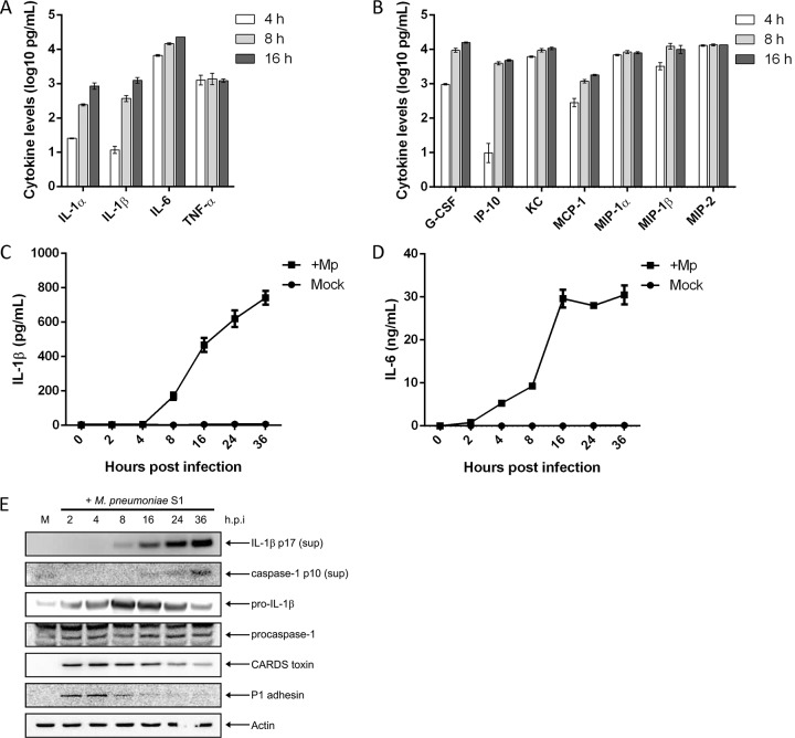
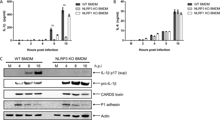
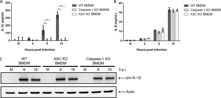
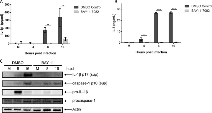
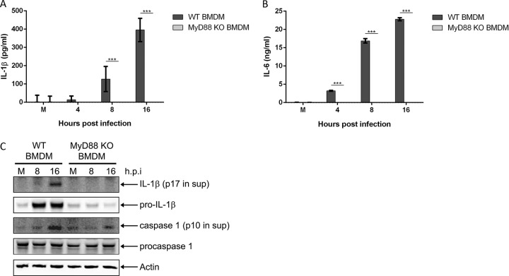
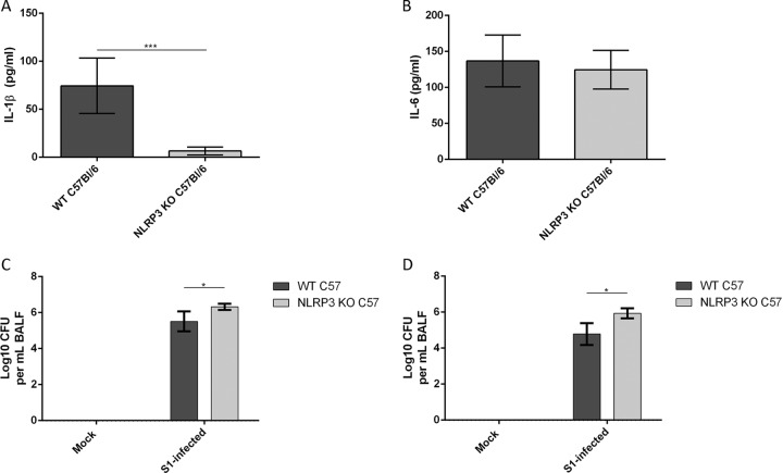
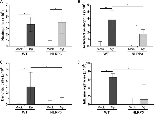
Mycoplasma pneumoniae is an atypical bacterial respiratory pathogen known to cause a range of airway inflammation and lung and extrapulmonary pathologies. We recently reported that an M. pneumoniae-derived ADP-ribosylating and vacuolating toxin called community-acquired respiratory distress syndrome (CARDS) toxin is capable of triggering NLRP3 (NLR-family, leucine-rich repeat protein 3) inflammasome activation and interleukin-1β (IL-1β) secretion in macrophages. However, it is unclear whether the NLRP3 inflammasome is important for the immune response during M. pneumoniae acute infection. In the current study, we utilized in vitro and in vivo models of M. pneumoniae infection to characterize the role of the NLRP3 inflammasome during acute infection. M. pneumoniae-infected macrophages deficient for inflammasome components NLRP3, ASC (apoptosis speck-like protein containing a caspase activation and recruitment domain), or caspase-1 failed to process and secrete IL-1β. The MyD88/NF-κB signaling pathway was found to be critical for proinflammatory gene expression in macrophages infected with M. pneumoniae C57BL/6 mice deficient for NLRP3 expression were unable to produce IL-1β in the airways during acute infection, and lack of this inflammatory response led to deficient immune cell activation and delayed bacterial clearance. These findings are the first to report the importance of the NLRP3 inflammasome in regulating the inflammatory response and influencing the progression of M. pneumoniae during acute infection.

Keywords: ADP-ribosylation; CARDS toxin; Mycoplasma pneumoniae; NLRP3; inflammasome; interleukin-1β.


Figures

FIG 1
FIG 1
Primary macrophages infected with M. pneumoniae produce proinflammatory and chemotactic cytokines. WT BMDMs were infected with M. pneumoniae at an MOI of 100 mycoplasmas per cell. At 4, 8, and 16 h postinfection, cell culture supernatants were collected and used in multiplex cytokine bead assays. Values for proinflammatory cytokines (A) and other cytokines and chemokines (B) were normalized by subtracting values of the uninfected control from those of the infected animals and are represented in log scale. Time course kinetics for IL-1β and IL-6 secretion are shown in panels C and D, respectively. (E) Immunoblot analysis of IL-1β p17 and caspase-1 p10 in cell culture supernatants (sup) and pro-IL-1β, procaspase-1, CARDS toxin, and P1 adhesin in cell lysates of WT BMDMs infected with M. pneumoniae. The mock-infected control is designated M. β-Actin served as the loading control. Data in panels A and B are representative of two independent experiments with similar results; data in panels C and D are representative of two independent experiments with similar results. h.p.i., hours postinfection; Mp, M. pneumoniae. All cytokine and chemokine levels are listed in Table S1 in the supplemental material.
FIG 2
FIG 2
NLRP3, but not NLRP1, is required for inflammasome activation and IL-1β secretion during M. pneumoniae infection. Cell culture supernatants from WT, NLRP3 KO, and NLRP1 KO BMDMs infected with M. pneumoniae were assayed by ELISA for IL-1β (A) and IL-6 (B) levels. (C) Immunoblot analysis of IL-1β p17 in supernatants and pro-IL-1β, CARDS toxin, and P1 adhesin in cell lysates of WT and NLRP3 KO BMDMs infected with M. pneumoniae. The mock-infected control is designated M. β-Actin served as the loading control. Data are representative of three independent experiments with similar results. **, P < 0.01. h.p.i., hours postinfection.
FIG 3
FIG 3
Caspase-1 and ASC are required for inflammasome activation and IL-1β processing during M. pneumoniae infection. Cell culture supernatants from WT, caspase-1 KO, and ASC KO BMDMs infected with M. pneumoniae were assayed by ELISA for IL-1β (A) and IL-6 (B) levels. (C) Immunoblot analysis for pro-IL-1β in cell lysates of WT, ASC KO, and caspase-1 KO BMDMs infected with M. pneumoniae. The mock-infected control is designated M. β-Actin served as the loading control. Data are representative of two independent experiments with similar results. **, P < 0.01. h.p.i., hours postinfection.
FIG 4
FIG 4
NF-κB signaling is required for expression of proinflammatory genes and inflammasome activation during M. pneumoniae infection. Cell culture supernatants from M. pneumoniae-infected WT BMDMs treated with the NF-κB inhibitor BAY11-7082 were assayed by ELISA for IL-1β (A) and IL-6 (B) levels. (C) Immunoblot analysis of IL-1β p17 and caspase-1 p10 in supernatants and pro-IL-1β and procaspase-1 in cell lysates of vehicle (DMSO-treated) or BAY11-7082-treated WT BMDMs infected with M. pneumoniae. The mock-infected control is designated M. β-Actin served as the loading control. Data are representative of three independent experiments with similar results. *, P < 0.05; **, P < 0.01; ***, P < 0.001. h.p.i., hours postinfection.
FIG 5
FIG 5
MyD88 signaling is required for expression of proinflammatory genes and inflammasome activation during M. pneumoniae infection. Cell culture supernatants from WT and MyD88 KO BMDMs infected with M. pneumoniae were assayed by ELISA for IL-1β (A) and IL-6 (B) levels. (C) Immunoblot analysis of IL-1β p17 and caspase-1 p10 in supernatants and pro-IL-1β and procaspase-1 in cell lysates of WT and MyD88 KO BMDMs infected with M. pneumoniae. The mock-infected control is designated M. β-Actin served as the loading control. Data are representative of three independent experiments with similar results. ***, P < 0.001. h.p.i., hours postinfection.
FIG 6
FIG 6
NLRP3 is required to elicit IL-1β production and to partly inhibit M. pneumoniae growth in vivo. WT and NLRP3 KO C57BL/6 mice were infected with M. pneumoniae (7 log10 CFU) for 2 days and 7 days. IL-1β (A) and IL-6 (B) levels in BALFs from 2-day-infected lungs were measured by ELISA. BALFs from mice at 2 days (C) and 7 days (D) postinfection were also analyzed for quantification of M. pneumoniae genomes by qPCR. *, P < 0.05; ***, P < 0.001.
FIG 7
FIG 7
Characterization of innate immune cell populations in lungs of WT and NLRP3 KO C57BL/6 mice infected with M. pneumoniae S1 for 2 days. Lungs from mock-infected or S1-infected WT and NLRP3 KO C57BL/6 mice were harvested, digested, and analyzed by fluorescence-activated cell sorting. Identified cell populations include total neutrophils (A), activated neutrophils (B), dendritic cells (C), and inflammatory macrophages (D). Cell populations in infected lungs were normalized to mock-infected cell populations by subtracting values of the uninfected controls from those of the infected animals. *, P < 0.05; **, P < 0.01.

References

    1. Guilbert TW, Denlinger LC. 2010. Role of infection in the development and exacerbation of asthma. Expert Rev Respir Med 4:71–83. doi: 10.1586/ers.09.60. - DOI - PMC - PubMed
    1. Jain S, Williams DJ, Arnold SR, Ampofo K, Bramley AM, Reed C, Stockmann C, Anderson EJ, Grijalva CG, Self WH, Zhu Y, Patel A, Hymas W, Chappell JD, Kaufman RA, Kan JH, Dansie D, Lenny N, Hillyard DR, Haynes LM, Levine M, Lindstrom S, Winchell JM, Katz JM, Erdman D, Schneider E, Hicks LA, Wunderink RG, Edwards KM, Pavia AT, McCullers JA, Finelli L, Team CES. 2015. Community-acquired pneumonia requiring hospitalization among U.S. children. N Engl J Med 372:835–845. doi: 10.1056/NEJMoa1405870. - DOI - PMC - PubMed
    1. Jain S, Self WH, Wunderink RG, Fakhran S, Balk R, Bramley AM, Reed C, Grijalva CG, Anderson EJ, Courtney DM, Chappell JD, Qi C, Hart EM, Carroll F, Trabue C, Donnelly HK, Williams DJ, Zhu Y, Arnold SR, Ampofo K, Waterer GW, Levine M, Lindstrom S, Winchell JM, Katz JM, Erdman D, Schneider E, Hicks LA, McCullers JA, Pavia AT, Edwards KM, Finelli L, CDC EPIC Study Team. 2015. Community-acquired pneumonia requiring hospitalization among U.S. adults. N Engl J Med 373:415–427. doi: 10.1056/NEJMoa1500245. - DOI - PMC - PubMed
    1. Waites KB, Talkington DF. 2004. Mycoplasma pneumoniae and its role as a human pathogen. Clin Microbiol Rev 17:697–728. doi: 10.1128/CMR.17.4.697-728.2004. - DOI - PMC - PubMed
    1. Yamada M, Buller R, Bledsoe S, Storch GA. 2012. Rising rates of macrolide-resistant Mycoplasma pneumoniae in the central United States. Pediatr Infect Dis J 31:409–400. doi: 10.1097/INF.0b013e318247f3e0. - DOI - PubMed

Publication types

MeSH terms

LinkOut - more resources