Review
. 2015 Nov 6;2(4):388-405.
doi: 10.3390/vetsci2040388.

Pathobiology of Hemangiosarcoma in Dogs: Research Advances and Future Perspectives

Affiliations
Review

Pathobiology of Hemangiosarcoma in Dogs: Research Advances and Future Perspectives

Jong-Hyuk Kim et al. Vet Sci. .

Abstract

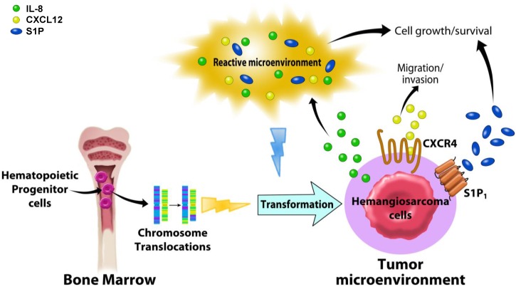
Hemangiosarcoma (HSA) is an aggressive and common cancer in dogs. While cutaneous masses are often treatable by tumor excision, visceral tumors are almost always incurable. Treatment advances for this disease have been limited due to a poor understanding of the overall tumor biology. Based upon its histological appearance, HSA has been presumed to originate from transformed endothelial cells; however, accumulating data now suggest a pluripotent bone marrow progenitor as the cell of origin for this disease. More recently, the identification of a novel subclassification of HSAs has provided a foundation to further our understanding of the cellular characteristics of HSA tumor cells, along with those of the cells comprising the tumor microenvironment. These discoveries hold promise for the development of new approaches to improve treatments for canine HSA, as well as to establish the utility of this disease as a spontaneous model to understand the pathogenesis and develop new treatments for vascular tumors of humans. In this review, we will provide a brief historical perspective and pathobiology of canine HSA, along with a focus on the recent advances in the molecular and cellular understanding of these tumors. In addition, future directions that should continue to improve our understanding of HSA pathogenesis will be discussed.

Keywords: animal model; dog; hemangiosarcoma; tumor microenvironment.


Conflict of interest statement

The authors declare no conflict of interest.

Figures

Figure 1
Figure 1
Histological appearance of canine hemangiosarcoma. (A) Capillary type; (B) Cavernous type; (C) Solid type. H&E stain. Magnification = 400×.
Figure 2
Figure 2
Expression of cell-surface determinants in canine HSA cell lines. One-dimensional overlay histograms show canine HSA cells stained with the indicated antibodies and analyzed by flow cytometry (gray). X-axes represent logarithmic fluorescence intensity and y-axes represent cell numbers (5000 to 10,000 events analyzed). Histograms are overlaid on corresponding negative controls using irrelevant antibodies (white). Data shown for DD-1 cells are from three experiments and represent more than seven experiments done. Data for Dal-4 cells represent more than four experiments done. Variability in antigen expression by these cells is described in the text. Reproduced with permission from [39].
Figure 3
Figure 3
Genome-wide expression analysis identified three molecular subtypes in hemangiosarcoma. Genes with variance >0.5 across 24 samples were used to generate principal component analyses. Samples were assigned to one of three groups by unsupervised clustering to identify genes with significantly different expression between groups (analysis of variance p < 0.001 and an average fold change >3 between groups). Reproduced with permission from [20].
Figure 4
Figure 4
Sphere cells serve as an in vitro model for HSA progenitor cells and express markers for endothelial and hematopoietic progenitors. (A) Flow cytometric analysis of expression of CD34, CD117, and CD133; (B) Graphical representation of the percentage of positive cells for each marker in the monolayer cells subtracted from the percentage of positive cells detected in the corresponding sphere cells. Results show a relative enrichment of the markers in sphere cells; (C) Heat map of 34 gene expression patterns significant in both the comparisons between the genes differently expressed in intact tumors and the genes differently expressed by spheres. For (C), each data set was independently mean centered. Reproduced with permission from [20].
Figure 5
Figure 5
Overview of the relationships between biological signaling molecules and the tumor microenvironment in canine hemangiosarcoma. Canine hemangiosarcoma is thought to originate from hematopoietic progenitor cells in the bone marrow. Chromosome translocations and a reactive microenvironment are suggested as potential genetic and biological events that may transform the hemangiosarcoma progenitor cells. Interleukin (IL)-8, produced by hemangiosarcoma cells, is thought to modulate the tumor microenvironment, promoting the growth and survival of tumor cells. CXCR4 and its ligand, CXCL12, found to be abundant in hemangiosarcoma tissue, transduce biological signaling, causing tumor cells to increase their motility to migrate and invade into the other sites for metastasis. Canine hemangiosarcoma cells that consume Sphingosine-1-phosphate (S1P) from the tumor microenvironment induce intracellular signaling through S1P receptor-1 (S1P1), increasing cell growth and survival. It is suggested that these chemokines and modified biolipids are key regulators for hemangiosarcoma behavior, and their signaling pathways are potential therapeutic targets.
Figure 6
Figure 6
Cytogenetic appearance of representative canine hemangiosarcoma cells. CXCR4, CXCR7, SLUG, and IL-8 genes were analyzed by fluorescence in situ hybridization (FISH) using BAC clones (CHORI-82). (Upper panel) Green and red spots indicate CXCR4 (CHORI-82-112B08) on CFA 19 and CXCR7 (CHORI-82-52B03) on CFA 25, respectively. (Bottom panel) Green and red spots indicate Slug (CHORI-82-130F20) on dog chromosome (CFA) 19 and IL-8 (CHORI-82-187A21) on CFA 13, respectively. Emma, JLU, and COSB = HSA cell lines.
Figure 7
Figure 7
Microscopic appearance of SB-HSA canine hemangiosarcoma xenografts. (A and B) Photomicrographs showing examples of tumors from two mice inoculated with 5 × 10⁶ SB-HSA cells stained with H & E; (C) Immunostaining of the tumor in a for Ki-67 using antibody MIB-1 (recognizes canine Ki-67); and (D) antibody TEC-3 (recognizes murine Ki-67). Magnification = 400×. Reproduced with permission from [22].

References

    1. Bergman P.J. Anticancer vaccines. Vet. Clin. North Am. Small Anim. Pract. 2007;37:1111–1119. - PubMed
    1. London C.A., Malpas P.B., Wood-Follis S.L., Boucher J.F., Rusk A.W., Rosenberg M.P., Henry C.J., Mitchener K.L., Klein M.K., Hintermeister J.G., et al. Multi-center, placebo-controlled, double-blind, randomized study of oral toceranib phosphate (Su11654), a receptor tyrosine kinase inhibitor, for the treatment of dogs with recurrent (either local or distant) mast cell tumor following surgical excision. Clin. Cancer Res. 2009;15:3856–3865. doi: 10.1158/1078-0432.CCR-08-1860. - DOI - PubMed
    1. Spirtas R., Beebe G., Baxter P., Dacey E., Faber M., Falk H., van Kaick G., Stafford J. Angiosarcoma as a model for comparative carcinogenesis (Letter to the Editor) Lancet. 1983 doi: 10.1016/S0140-6736(83)90417-8. - DOI - PubMed
    1. Falk H., Herbert J., Crowley S., Ishak K.G., Thomas L.B., Popper H., Caldwell G.G. Epidemiology of hepatic angiosarcoma in the United States: 1964–1974. Environ. Health Perspect. 1981;41:107–113. doi: 10.1289/ehp.8141107. - DOI - PMC - PubMed
    1. Lieberman L.L. Malignant hemangioendothelioma of the canine heart. J. Am. Vet. Med. Assoc. 1955;126:296. - PubMed

LinkOut - more resources