. 2017 Oct 23;7(1):13754.
doi: 10.1038/s41598-017-14079-4.

Canonical NFκB signaling in myeloid cells is required for the glioblastoma growth

Affiliations

Canonical NFκB signaling in myeloid cells is required for the glioblastoma growth

B R Achyut et al. Sci Rep. .

Abstract

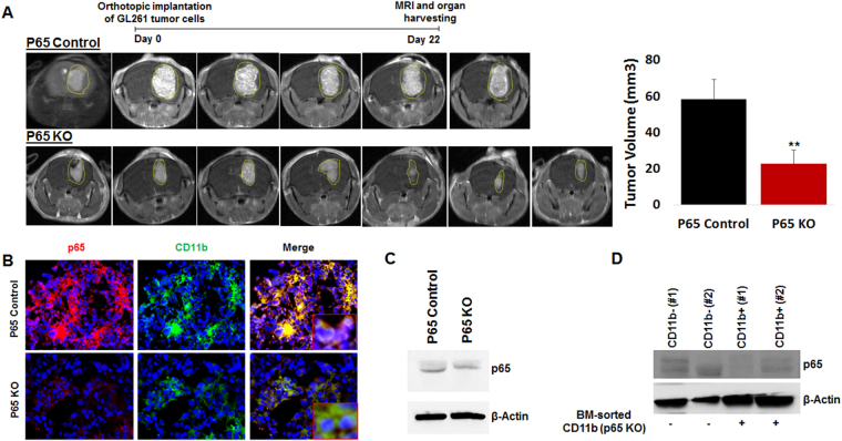
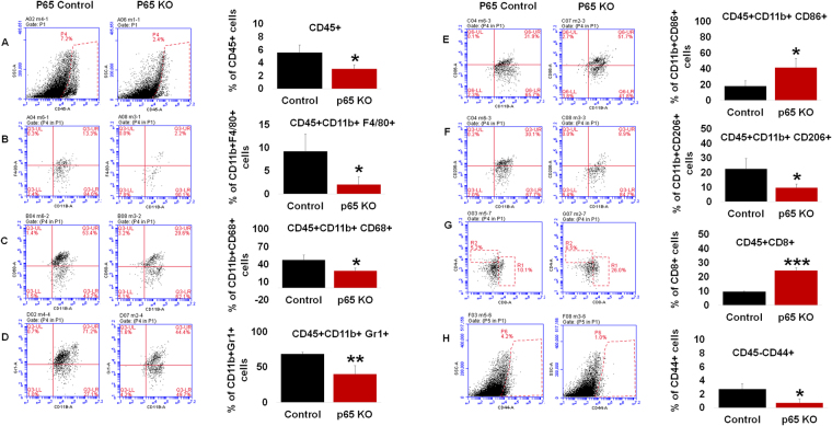
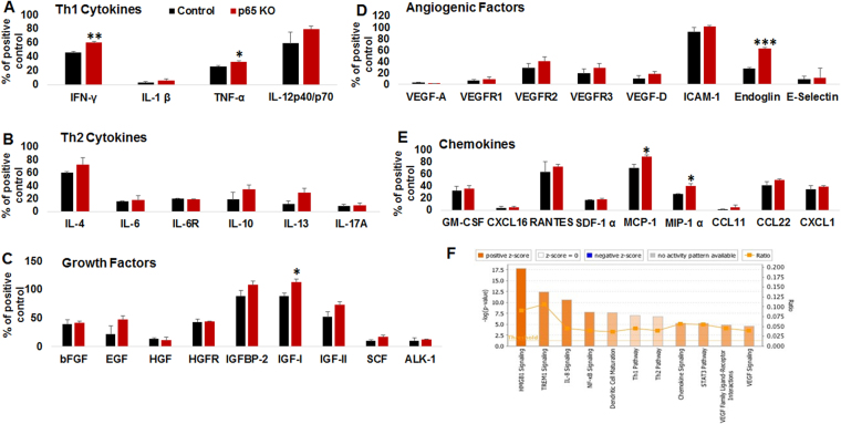
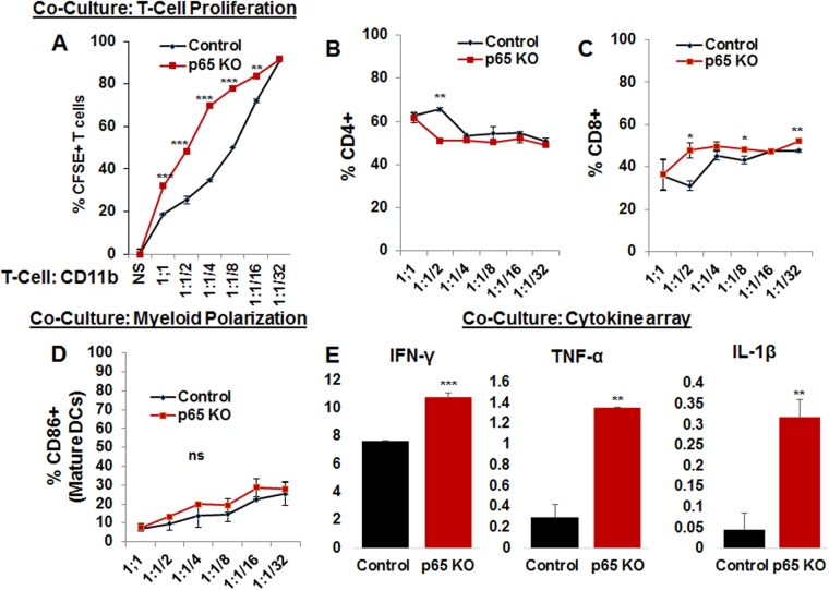
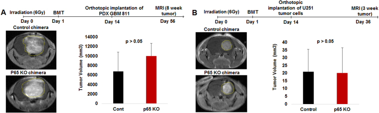
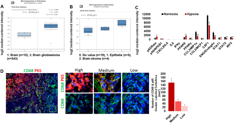
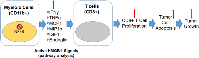
Tumor development and therapeutic resistance are linked with tumor-associated macrophage (TAM) and myeloid-derived suppressor cell (MDSC) infiltration in tumors via chemokine axis. Chemokine expression, which determines the pro or anti-inflammatory status of myeloid cells, are partly regulated by the nuclear factor-kappa B (NF-κB) pathway. Here, we identified that conditional deletion of canonical NF-κB signaling (p65) in myeloid cells inhibited syngeneic glioblastoma (GBM) through decreased CD45 infiltration in tumors, as characterized by decreased TAMs (CD206+) and MDSCs (Gr1+ CD11b+), increased dendritic cells (CD86+) and cytotoxic T cells (CD8+) in the p65 knockout (KO) mice. Proinflammatory cytokines (IFNγ, MCP1, MIP1α, and TNFα) and myeloid differentiation factor (Endoglin) were increased in myeloid cells from p65 KO tumor, which demonstrated an influence on CD8+T cell proliferation. In contrast, p65KO athymic chimeric mice with human GBM, failed to inhibit tumor growth, confirming the contribution of T cells in an immune competent model. The analysis of human datasets and GBM tumors revealed higher expression of p65 in GBM-associated CD68+ macrophages compared to neighboring stroma. Thus, canonical NF-κB signaling has an anti-inflammatory role and is required for macrophage polarization, immune suppression, and GBM growth. Combining an NF-κB inhibitor with standard therapy could improve antitumor immunity in GBM.


Conflict of interest statement

The authors declare that they have no competing interests.

Figures

Figure 1
Figure 1
Myeloid-specific deletion of NF-κB signaling decreases syngeneic GBM growth. (A) Syngeneic GL261 GBM tumors were developed in p65 control and p65 KO mice. MRI data showing significantly decreased GBM tumors in p65KO mice compared to controls. (B) Tissue-based immunofluorescence images showing decreased p65 expression in myeloid cells from p65 KO tumors compared with control tumors. (C) Brain sorted microglia displayed comparable p65 expression in both control and p65 KO mice. (D) Western blot data showing p65 expression in bone marrow sorted CD11b+ and CD11b− cells. The p65 KO mice, which were identified for incomplete p65 loss at the protein level in CD11b+ cells (e.g. mouse #2), were excluded from the study. Shown is one of the two experiments performed. Quantitative data is expressed in mean ± SD. **P < 0.01.
Figure 2
Figure 2
Myeloid-specific deletion of NF-κB signaling polarizes microenvironment: Flow cytometry data showing (A) Myeloid-specific deletion of NF-κB signaling resulted in decreased infiltration of bone marrow-derived CD45+ leukocytes in the TME compared with the control mice. (B) CD45+ leukocytes were characterized as decreased common macrophages (F4/80+), (C) tumor associated macrophages (CD68+), (D) decreased MDSCs (Gr1+ CD11b+), (E) increased M1 macrophages (CD86+), (F) decreased M2 macrophages (CD206+ or mannose receptor), (G) increased CD8+T cells in the p65 KO tumors compared to control tumors. (H) Flow cytometry data showing decreased CD45− and CD44+, mesenchymal stem cell (MSCs)-like cells in the p65 KO tumors compared to control tumors. Shown is one of the two experiments performed. Quantitative data is expressed in mean ± SD. *P < 0.05, **P < 0.01 and ***P < 0.001.
Figure 3
Figure 3
Myeloid-specific deletion of NF-κB signaling displayed anti-tumor secretome: Membrane-based cytokine array data showing (A) increased expression of IFN-γ and TNF-α (Th1 cytokine), (B) no change in Th2 cytokines, (C) increased expression of IGF-1 (growth factor), (D) increased expression of endoglin (myeloid maturation factor), and (E) increased expression of MCP-1 and MIP-1α (chemokines) in p65 deleted CD11b+ myeloid cells compared to p65 control myeloid cells. (F) web-based pathway analysis discovered that significantly altered factors predominantly belonged to the HMGB1 signaling pathways. Interestingly, altered factors also found to be associated with top 4 pathways TREM1 > IL8 > NF-κB > dendritic cell maturation. Shown is one of the two experiments performed. Quantitative data is expressed in mean ± SD. *P < 0.05, **P < 0.01 and ***P < 0.001.
Figure 4
Figure 4
Myeloid-specific NF-κB signaling inhibits T cell proliferation: In vitro data showing co-culture experiments with sorted and CFSE labeled CD3+T cells and tumor isolated CD11b+ from p65KO or p65 control mice. (A) flow cytometry data showing that tumor sorted CD11b+ cells from p65 KO mice displayed higher T cell proliferation at 1:1, 1:1/2, 1:1/4, 1:1/8 and 1:1/16 compared to control CD11b+ cells. (B) CD4 proliferation was decreased at the 1:1/2 co-culture and (C) CD8 proliferation was increased at the 1:1/2, 1:1/8 and 1:1/32 co-cultures in the p65KO group compared to p65 control group. (D) flow data showing an increasing trend of CD86+ mature DCs in p65KO co-cultures compared to p65 controls. (E) analysis of co-culture supernatant using membrane-based cytokine array depicted higher levels of IFN-γ, TNF-α, and IL1-β (Th1 cytokines) in p65KO co-culture (1:1) compared to p65 controls. Shown is one of the two experiments performed. Quantitative data is expressed in mean ± SD. *P < 0.05, **P < 0.01 and ***P < 0.001.
Figure 5
Figure 5
p65 KO chimera failed to inhibit human GBM growth in immune-deficient host. (A and B) The p65 KO and control recipient mice bone marrow was isolated and transplanted into irradiated donor nude mice. Once mice achieved 70% engraftment (chimera) in 2 weeks, U251 or PDX GBM811 cells were orthotopically implanted and followed-up for MRI at the 3-week or 8-week protocol, respectively. MRI data showing p65KO chimera failed to decrease the U251 or PDX GBM811 human tumor growth. Shown is one of the two experiments performed. Quantitative data is expressed in mean ± SD.
Figure 6
Figure 6
Elevated stromal NF-κB signaling in human brain tumors. (A) The TCGA brain data showing increased p65 expression in GBM compared to normal brain. (B) Analysis of tumor and stroma in Albino brain dataset identified increased p65 expression in stroma compared to tumor compartment. (C) GEO dataset (GSE4630) showing hypoxia-induced p65 mRNA expression associated mediators involved in inflammation and myeloid differentiation in hypoxic human macrophages compared to macrophages cultured in normoxia. (D) Analysis of paraffin fixed GBM brain tissues identified heterogeneous p65 protein expression pattern in CD68+ myeloid cells.
Figure 7
Figure 7
Schematic showing increased expression of pro-inflammatory HMGB1 signals in canonical NF-κB deleted myeloid cells modulate proliferation of CD8+T cells and increase apoptosis in the TME to limit GBM growth.

References

    1. Olar A, Aldape KD. Using the molecular classification of glioblastoma to inform personalized treatment. The Journal of pathology. 2014;232:165–177. doi: 10.1002/path.4282. - DOI - PMC - PubMed
    1. Brat DJ, Van Meir EG. Vaso-occlusive and prothrombotic mechanisms associated with tumor hypoxia, necrosis, and accelerated growth in glioblastoma. Laboratory investigation; a journal of technical methods and pathology. 2004;84:397–405. doi: 10.1038/labinvest.3700070. - DOI - PubMed
    1. Stupp R, et al. Radiotherapy plus concomitant and adjuvant temozolomide for glioblastoma. N Engl J Med. 2005;352:987–996. doi: 10.1056/NEJMoa043330. - DOI - PubMed
    1. Lahmar Q, et al. Tissue-resident versus monocyte-derived macrophages in the tumor microenvironment. Biochimica et biophysica acta. 2016;1865:23–34. - PubMed
    1. Morantz RA, Wood GW, Foster M, Clark M, Gollahon K. Macrophages in experimental and human brain tumors. Part 2: studies of the macrophage content of human brain tumors. Journal of neurosurgery. 1979;50:305–311. doi: 10.3171/jns.1979.50.3.0305. - DOI - PubMed

Publication types

MeSH terms