Review
. 2018 Jan;16(1):19-31.
doi: 10.1038/nrmicro.2017.107. Epub 2017 Oct 3.

Development and regulation of single- and multi-species Candida albicans biofilms

Affiliations
Review

Development and regulation of single- and multi-species Candida albicans biofilms

Matthew B Lohse et al. Nat Rev Microbiol. 2018 Jan.

Abstract

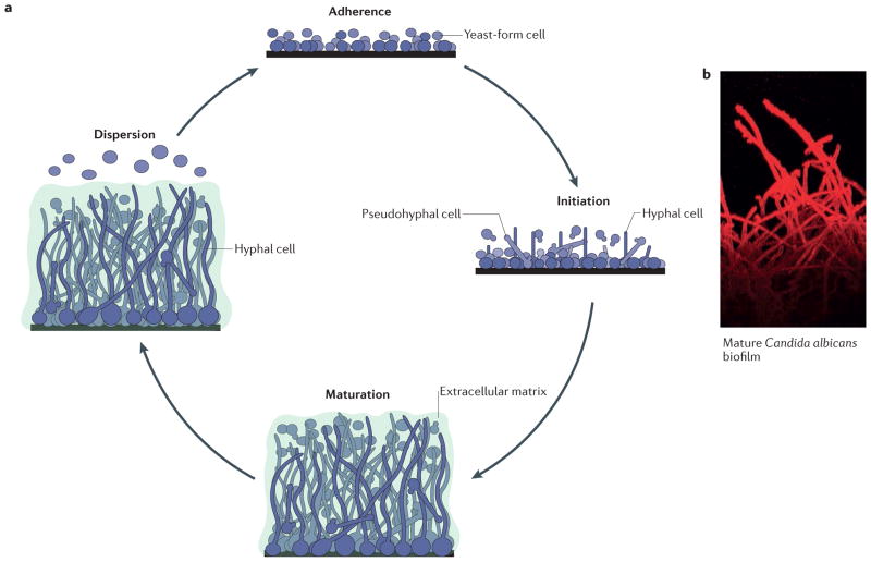
Candida albicans is among the most prevalent fungal species of the human microbiota and asymptomatically colonizes healthy individuals. However, it is also an opportunistic pathogen that can cause severe, and often fatal, bloodstream infections. The medical impact of C. albicans typically depends on its ability to form biofilms, which are closely packed communities of cells that attach to surfaces, such as tissues and implanted medical devices. In this Review, we provide an overview of the processes involved in the formation of C. albicans biofilms and discuss the core transcriptional network that regulates biofilm development. We also consider some of the advantages that biofilms provide to C. albicans in comparison with planktonic growth and explore polymicrobial biofilms that are formed by C. albicans and certain bacterial species.


Conflict of interest statement

Competing interests statement

The authors declare competing interests: see Web version for details.

Figures

Figure 1
Figure 1. Formation of Candida albicans biofilms
a | The formation of Candida albicans biofilms has been divided into four major stages: adherence of round yeast-form cells to a surface; initiation of biofilm formation, during which the cells adhered to the surface form a basal layer that contains yeast-form, pseudohyphal and hyphal cells (also known as the proliferation stage); maturation into a complex, structured biofilm, in which cells are encased in the extracellular matrix; and dispersion of yeast-form cells from the biofilm to seed new sites. b | A confocal laser-scanning microscopy image of the side view of a mature C. albicans biofilm is shown, with the long hyphal cells clearly visible. The dye (concanavalin A–Alexa Fluor 594 conjugate) used for imaging does not penetrate to the bottom of the biofilm; hence, the yeast-form cells attached to the solid surface are not readily visible. The extracellular matrix is also not visible, as it does not bind the dye. Part a modified with permission from the Annual Review of Microbiology, Volume 69 © 2015 by Annual Reviews, http://www.annualreviews.org.
Figure 2
Figure 2. The core transcriptional network controlling biofilm formation in Candida albicans
More than 50 transcriptional regulators have been linked to the formation of Candida albicans biofilms. The proteins depicted are a ‘core’ set of nine regulators (Ndt80, Bcr1, Rfx2, Flo8, Rob1, Brg1, Gal4, Tec1 and Efg1) that is required for biofilm development. Autoregulation is indicated by dotted arrows, direct binding interactions between two regulators that each regulate the activity of the other are indicated by double-headed dark grey arrows, and direct binding interactions where one regulator controls another regulator are indicated by single-headed light grey arrows. The indicated interactions are based on previously reported chromatin immunoprecipitation data,.
Figure 3
Figure 3. Overview of Candida albicans biofilm antifungal drug resistance
Characteristics of Candida albicans biofilms that contribute to resistance to antifungal drugs. C. albicans biofilms are complex structures that contain round, yeast-form cells, pseudohyphal cells and hyphal cells (shown in blue) that are encased in an extracellular matrix (shown in green). The extracellular matrix functions as a physical barrier to antifungal drugs. Cells within the biofilm also exhibit increased cell density, increased stress response and decreased metabolic activity, which all contribute to antifungal drug resistance. A minority cell population, called ‘persister’ cells (shown in orange), can exist in the basal layer of the biofilm. Persister cells are non-dividing cells with decreased metabolic activities, making them highly resistant to antimicrobial drugs and likely to seed new biofilm infections after drug treatment. Efflux pumps export drug molecules from the inside the cell to the environment. Expression of these efflux pumps is highly upregulated in C. albicans biofilms, even in the absence of antifungal drugs, which contributes to the overall drug-resistant nature of biofilms.
Figure 4
Figure 4. Multi-species biofilm formation
a | Bacterial species most frequently isolated with Candida albicans from specific niches of the human body, including the oral cavity, lungs, gastrointestinal tract, vulvovaginal region and skin are listed. Infections resulting from the presence of a burn wound or an implanted medical device are often sites for bacterial–fungal infections; bacterial species most commonly isolated from these infections are listed. b–f | Common ways in which C. albicans interacts with various bacterial species in the context of a biofilm.b | Bacterial cells (Gram-positive cocci and bacilli are shown in red with a black outline; Gram-negative bacilli are shown in red with a red outline) can directly bind to C. albicans hyphal cells (shown in blue). c | C. albicans–bacterial interactions can be synergistic; for example, aC. albicans biofilm can physically protect bacteria from antimicrobial agents (shown in yellow) or can protect anaerobic bacteria from high oxygen concentrations by providing a low oxygen niche within the depths of the biofilm.d | Signalling molecules produced by bacterial species and C. albicans enable inter-kingdom communication within mixed-species biofilms. For example, in C. albicansPseudomonas aeruginosa mixed-species biofilms, the P. aeruginosa-secreted signalling molecule 3-oxo-C12-homoserine lactone (shown in orange) can influence the behaviour ofC. albicans, and the C. albicans secreted signalling molecule farnesol (shown in purple) can influence the behaviour of P. aeruginosa. e | C. albicans–bacterial interactions can be antagonistic; for example, acid produced byLactobacillus spp. results in a lower local pH, which inhibits C. albicans hyphal formation. f | Cells within mixed-species biofilms can also exchange nutrients. For example, cells can share nutrients (shown in green), or one species can utilize available nutrients (blue and orange) and in turn produce nutrients (yellow and purple) needed by the other species. S. gordonii, Streptococcus gordonii; S. mutans, Streptococcus mutans; S. salivaris, Streptococcus salivaris.

References

    1. Nobile CJ, Johnson AD. Candida albicans biofilms and human disease. Annu Rev Microbiol. 2015;69:71–92. - PMC - PubMed
    1. Davey ME, O’Toole GA. Microbial biofilms: from ecology to molecular genetics. Microbiol Mol Biol Rev. 2000;64:847–867. - PMC - PubMed
    1. Kolter R, Greenberg EP. Microbial sciences: The superficial life of microbes. Nature. 2006;441:300–302. - PubMed
    1. Hall-Stoodley L, Costerton JW, Stoodley P. Bacterial biofilms: from the natural environment to infectious diseases. Nat Rev Microbiol. 2004;2:95–108. - PubMed
    1. Wolcott R, Costerton JW, Raoult D, Culter SJ. The polymicrobial nature of biofilm infection. Clin Microbiol Infect. 2013;19:107–112. - PubMed

MeSH terms

Substances