. 2017 Oct 9:11:313.
doi: 10.3389/fncel.2017.00313. eCollection 2017.

Obesity Increases Mitogen-Activated Protein Kinase Phosphatase-3 Levels in the Hypothalamus of Mice

Affiliations

Obesity Increases Mitogen-Activated Protein Kinase Phosphatase-3 Levels in the Hypothalamus of Mice

Bárbara de A Rodrigues et al. Front Cell Neurosci. .

Abstract

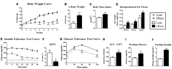
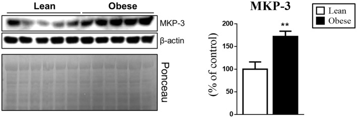
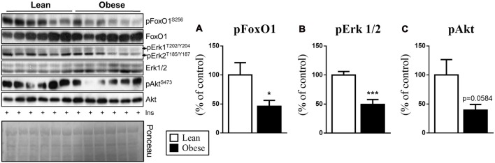
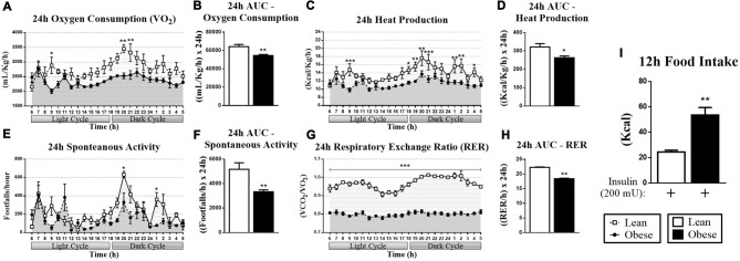
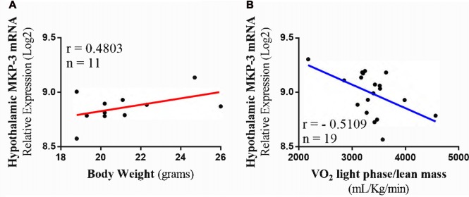
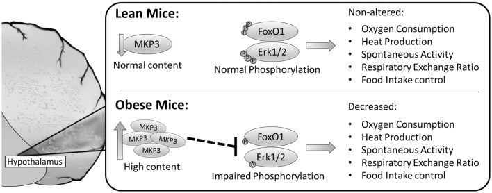
Mitogen-activated Protein Kinase Phosphatase 3 (MKP-3) has been involved in the negative regulation of insulin signaling. The absence of MKP-3 is also associated with reduced adiposity, increased energy expenditure and improved insulin sensitivity. The MKP-3 is known as the main Erk1/2 phosphatase and FoxO1 activator, which has repercussions on the gluconeogenesis pathway and hyperglycemia in obese mice. Recently, we showed that MKP-3 overexpression decreases FoxO1 phosphorylation in the hypothalamus of lean mice. However, the hypothalamic interaction between MKP-3 and FoxO1 during obesity was not investigated yet. Here, the MKP-3 expression and the effects on food intake and energy expenditure, were investigated in high-fat diet-induced obese mice. The results indicate that obesity in mice increased the MKP-3 protein content in the hypothalamus. This hypothalamic upregulation led to an increase of food intake, adiposity, and body weight. Furthermore, the obese mice with increased MKP-3 showed an insulin signaling impairment with reduction of insulin-induced FoxO1 and Erk1/2 phosphorylation in the hypothalamus. Moreover, a bioinformatics analysis of data demonstrated that hypothalamic MKP-3 mRNA levels were positively correlated with body weight and negatively correlated to oxygen consumption (VO2) in BXD mice. Taken together, our study reports that obesity is associated with increased protein levels of hypothalamic MKP-3, which is related to the reduction of FoxO1 and Erk1/2 phosphorylation in the hypothalamus as well as to an increase in body weight and a reduction in energy expenditure.

Keywords: MKP-3; food intake; hypothalamus; insulin; obesity.


Figures

Figure 1
Figure 1
Metabolic and physiological parameters of lean and obese animals. (A) Body mass curve during 8 weeks. (B) Δ Body mass between the first and eighth week of treatment. (C) Body mass index (BMI; g/cm2). (D) The retroperitoneal, mesenteric and epididymal fat mass weight/body weight. (E) Insulin Tolerance Test (ITT) curve. (F) ITT KITT curve. (G) Glucose Tolerance Test (GTT) curve. (H) Area under the curve (AUC) of the ITT. (I) Fasting glucose. (J) Fasting insulin. The bars and lines in the graphs represent the mean and standard error of the mean (SEM; n = 6–10). *p < 0.05 vs. Lean group. **p < 0.01 vs. Lean group. ***p < 0.001 vs. Lean group.
Figure 2
Figure 2
Increased hypothalamic Mitogen-activated Protein Kinase Phosphatase 3 (MKP-3) content in obese mice. MKP-3 protein content in the hypothalamus of lean and obese mice. The bars in the graphs represent the mean and SEM (n = 5). **p < 0.01 vs. Lean group.
Figure 3
Figure 3
Phosphorylation of the proteins involved with the MKP-3 phosphatase activity (pFoxO1 and pErk1/2) after insulin stimulus. (A) FoxO1 phosphorylation (S256) normalized by its total content. (B) Erk1/2 phosphorylation (T202/Y204/T185/Y187) normalized by its total content. (C) Akt phosphorylation (S473) normalized by its total content. Related to the total content of FoxO1, Erk and Akt were used as a control, only Erk1/2 showed a significant increase compared to the CTL group. The bars in the graphs represent the mean and SEM (n = 6). *p < 0.05 vs. Lean group. ***p < 0.001 vs. Lean group.
Figure 4
Figure 4
Respirometry and dietary intake data of lean and obese mice. (A) Oxygen consumption (VO2) during 24 h. (B) AUC for the VO2 during 24 h. (C) Heat production measured during 24 h. (D) AUC for the heat production during 24 h. (E) Spontaneous activity during 24 h. (F) AUC for the spontaneous activity during 24 h. (G) Respiratory exchange ratio (RER) during 24 h. (H) AUC for the RER during 24 h. (I) Cumulative food intake of 12 h after insulin stimulation. The bars and lines in the graphs represent the mean and SEM (n = 6). *p < 0.05 vs. Lean group. **p < 0.01 vs. Lean group. ***p < 0.001 vs. Lean group.
Figure 5
Figure 5
Transcriptome and phenotype bioinformatics analysis. (A) Positive Pearson’s correlation between hypothalamic MKP-3 mRNA in lean mice and body weight (n = 11). (B) Negative Pearson’s correlation between hypothalamic MKP-3 mRNA and oxygen consumption (VO2) in light phase (n = 19).
Figure 6
Figure 6
Schematic model summarizing the possible molecular mechanisms by which MKP-3 led to food intake increase and thermogenesis decrease. The obese mice presented high content of MKP-3 in the hypothalamus, and decreased phosphorylation of FoxO1 and Erk1/2 leading to decreased oxygen consumption, heat production, spontaneous activity, RER and increased food intake.

Similar articles

Cited by

References

    1. Andreux P. A., Williams E. G., Koutnikova H., Houtkooper R. H., Champy M.-F., Henry H., et al. . (2012). Systems genetics of metabolism: the use of the BXD murine reference panel for multiscalar integration of traits. Cell 150, 1287–1299. 10.1016/j.cell.2012.08.012 - DOI - PMC - PubMed
    1. Aoki M., Jiang H., Vogt P. K. (2004). Proteasomal degradation of the FoxO1 transcriptional regulator in cells transformed by the P3k and Akt oncoproteins. Proc. Natl. Acad. Sci. U S A 101, 13613–13617. 10.1073/pnas.0405454101 - DOI - PMC - PubMed
    1. Arruda A. P., Milanski M., Coope A., Torsoni A. S., Ropelle E., Carvalho D. P., et al. . (2011). Low-grade hypothalamic inflammation leads to defective thermogenesis, insulin resistance, and impaired insulin secretion. Endocrinology 152, 1314–1326. 10.1210/en.2010-0659 - DOI - PubMed
    1. Botezelli J. D., Coope A., Ghezzi A. C., Cambri L. T., Moura L. P., Scariot P. P. M., et al. . (2016). Strength training prevents hyperinsulinemia, insulin resistance, and inflammation independent of weight loss in fructose-fed animals. Sci. Rep. 6:31106. 10.1038/srep31106 - DOI - PMC - PubMed
    1. Cintra D. E., Ropelle E. R., Moraes J. C., Pauli J. R., Morari J., de Souza C. T., et al. . (2012). Unsaturated fatty acids revert diet-induced hypothalamic inflammation in obesity. PLoS One 7:e30571. 10.1371/journal.pone.0030571 - DOI - PMC - PubMed