. 2017 Oct 19:3:25.
doi: 10.1038/s41522-017-0035-0. eCollection 2017.

Staphylococcus aureus interaction with Pseudomonas aeruginosa biofilm enhances tobramycin resistance

Affiliations

Staphylococcus aureus interaction with Pseudomonas aeruginosa biofilm enhances tobramycin resistance

T Beaudoin et al. NPJ Biofilms Microbiomes. .

Abstract

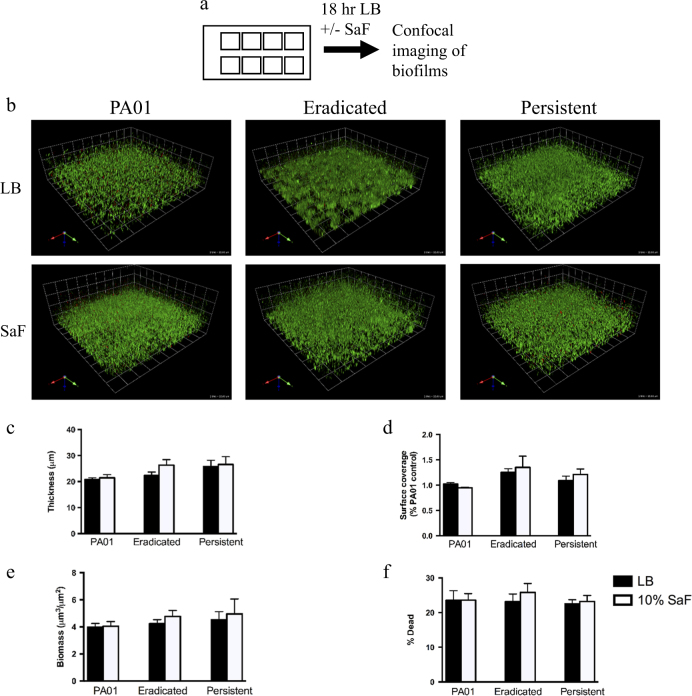
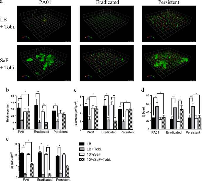
Antimicrobial resistance is a significant threat to the treatment of infectious disease. Multiple mechanisms of resistance to different classes of antibiotics have been identified and well-studied. However, these mechanisms are studied with bacteria in isolation, whereas often, infections have a polymicrobial basis. Using a biofilm slide chamber model, we visualized the formation and development of clinical Pseudomonas aeruginosa biofilms in the presence of secreted Staphylococcus aureus exoproducts, two bacteria that commonly co-infect pediatric patients with cystic fibrosis. We showed that, over time, certain isolates of P. aeruginosa can form different biofilm architecture in the presence of S. aureus exoproducts. We further determined that this interaction was dependent on Psl produced by P. aeruginosa and staphylococcal protein A from S. aureus. Importantly, we identified a mechanism of antibiotic resistance to tobramycin that is dependent on the polymicrobial interactions between these two bacteria. This interaction occurred in isolates of P. aeruginosa recovered from children with cystic fibrosis who failed to clear P. aeruginosa following inhaled tobramycin treatment.


Conflict of interest statement

The authors declare that they have no competing financial interests.

Figures

Fig. 1
Fig. 1
Staphylococcus aureus filtrates (SaF) do not affect initial attachment of Pseudomonas aeruginosa to borosilicate slide chambers. a Schematic representation of how SaF biofilm formation was assessed. P. aeruginosa was grown overnight in liquid culture prior to seeding in 8-well slide chambers. After 6 h of attachment, media was removed and replaced with fresh media. b Representative images of P. aeruginosa biofilms grown for 24 h in the presence of 10% (v/v) SaF in LB. Green cells: live; red cells: dead. cf Image analysis of biofilm thickness c, surface coverage d, biomass e and % dead cells f for given biofilms. Biofilms were grown as described above in the presence of LB alone (black bars) or 10%SaF (white bars) prior to image acquisition and analysis with COMSTAT. N = 7 eradicated isolates and N = 7 persistent isolates. The mean of PA01 was generated from seven biological replicates. All means are plotted with standard deviation. Statistics were performed using non-parametric one-way ANOVA (Kruskal–Wallis) with Dunn’s post-test for multiple comparisons
Fig. 2
Fig. 2
Staphylococcus aureus filtrates (SaF) differentially affects Pseudomonas aeruginosa development in a slide chamber model. a Schematic representation of how SaF biofilm formation was assessed. b Representative images of P. aeruginosa biofilms grown for 24 h in the presence of 10% (v/v) SaF in LB. Green cells: live; red cells: dead. cf Image analysis of biofilm thickness c, surface coverage d, biomass e and % dead cells f for PAO1, eradicated (N = 7) and persistent (N = 7) P. aeruginosa isolates biofilms. Biofilms were grown as described above in the presence of LB alone (black bars) or 10%SaF (white bars) prior to image acquisition and analysis with COMSTAT. The mean of PA01 was generated from seven biological replicates. g CFU/cm2 of biofilms grown with or without SaF. h Proportion of CFU of unattached bacteria (in media) to attached bacteria (on surface) in LB or 10% SaF. All means are plotted with standard deviation. Statistics were performed using non-parametric one-way ANOVA (Kruskal–Wallis) with Dunn’s post-test for multiple comparisons. **p < 0.001
Fig. 3
Fig. 3
Staphylococcus aureus filtrates (SaF) leads to increased tobramycin resistance in P. aeruginosa biofilms formed from persistent isolates A static biofilm slide chamber method was used to asses the effect of S. aureus filtrates (SaF) on PAO1, eradicated (N = 7) and persistent (N = 7) P. aeruginosa antibiotic tolerance. a Representative images of P. aeruginosa biofilms grown for 24 h in LB followed by exposure to 10% SaF and or 1000 µg/mL of tobramycin for an additional 24 h prior to staining. Green cells: live; red cells: dead. b–e Image analysis of biofilm thickness b, biomass c % dead cells d and CFU counts/cm2 e for given biofilms. Biofilms were grown as described above in the presence of LB alone (black bars), LB + tobramycin (gray bars), 10%SaF (white bars) or 10%SaF + tobramycin (checkered bars) prior to image acquisition and analysis with COMSTAT. Each biological replicate consisted of analyzing six images per isolate. The mean of PA01 was generated from seven biological replicates. All means are plotted with standard deviation. Statistics were performed using non-parametric one-way ANOVA (Kruskal–Wallis) with Dunn’s post-test for multiple comparisons. **p < 0.001, *p < 0.05
Fig. 4
Fig. 4
Staphylococcal protein A leads to aggregation in Pseudomonas aeruginosa biofilms and increases antibiotic tolerance. a Persistent isolate (PA580) grown in LB for 24 h followed by 24 h of growth in 10% SaF that had undergone various manipulations and analyzed for % surface coverage as a marker for aggregation. H-SaF; Heat-inactivated SaF, Prot. K: Proteinase K (0.5 mg/mL). Images from n = 3 independent experiments were analyzed. Mean values are plotted with standard deviation b FITC-SpA assay of Pseudomonas aeruginosa isolates grown for 24 h in 96-well microtiter plate in LB media alone. After 24 h of growth, cells were spun down and suspended in 50 µg/mL of FITC-labeled SpA. For eradicated (n = 33) and persistent isolate (n = 13), each dot represents the mean for an individual isolate obtained from 2 biological replicates. For each biological replicate, all isolates were performed in quadruplicate. For PAO1 (n = 9) and PAO1ΔpslBCD (n = 9), each dot represents a biological replicate. Mean values are plotted with standard deviation. c Eradicated or persistent isolates were grown for 24 h in LB followed by 24 h in LB + /− 50 µg/mL SpA or SaF with SpA removed via IgG sepharose column. N = 3 eradicated or persistent isolates performed in 3 independent experiments. Isolates were grown in LB alone (black bars), 10% SaF (white bars), 50 µg/mL SpA (dark gray bars) or SaF with SpA removed (hashed bars). Images were analyzed with COMSTAT and mean surface coverage is shown. de Eradicated isolates or persistent isolates were grown for 24 h in LB followed by 24 h in LB + /− 50 µg/mL SpA with or without tobramycin. N = 3 eradicated or persistent isolates performed in 3 independent experiments. Average biomass d and % dead e of biofilms as described above in LB (Black bars), LB + tobramycin (White bars), SpA (dark gray bars) or SpA + tobramycin (hatched bars). All means are plotted with standard deviation. Statistics were performed using non-parametric one-way ANOVA (Kruskal–Wallis) with Dunn’s post-test for multiple comparisons. **p < 0.001, *p < 0.05
Fig. 5
Fig. 5
Binding of Staphylococcal protein A to Psl exopolysaccharide in Pseudomonas aeruginosa biofilms leads to increased tobramycin resistance. a–b Total volume of Psl stain a and SpA b in biofilm as determined by voxel counts using Volocity software. N = 4 eradicated and N = 4 persistent isolates performed n = 3 times. Mean values are plotted with standard deviation. c–d Biofilms were grown for 48 h in the presence of or absence of SaF or SpA. Following the initial 24 h period, media was removed and replaced with fresh media with or without SaF or Spa and with or without tobramycin. Images were taken and analyzed for average biomass c and % dead d of biofilms as described. Black bars represent biofilms without tobramycin, white bars represent biofilms grown in presence of tobramycin. Experiment was performed n = 3 times. Mean values are plotted with standard error of the mean. Statistics were performed using non-parametric one-way ANOVA (Kruskal–Wallis) with Dunn’s post-test for multiple comparisons. **p < 0.001, *p < 0.05 compared to LB control
Fig. 6
Fig. 6
Exogenous exopolysaccharides derived from PA01 or persistent isolates, and in the presence of SpA, can protect eradicated isolates biofilms from tobramycin. a Representative images of eradicated isolate grown in the presence of exopolysaccharides produced from different isolates of Pseudomonas aeruginosa. Eradicated isolates were grown for 24 h in media alone, followed by 24 h in the presence of 10% extracted biofilm matrix from an eradicated isolate with 50 µg/mL SpA (Eradic. Exo + SpA) or from biofilm matrix obtained from a persistent isolate in the presence or absence of 50 µg/mL SpA for 2 h. Following 2 h treatment, tobramycin was added to a final concentration of 1000 µg/mL and biofilm was allowed to grow for an additional 24 h before imaging. Images were obtained and analyzed for, biomass b and % dead c of biofilms without (Black bars) tobramycin or in the presence (White bars) of tobramycin. N = 3 Eradicated isolates performed n = 3 times. Mean values are plotted with standard deviation. Statistics were performed using non-parametric one-way ANOVA (Kruskal–Wallis) with Dunn’s post-test for multiple comparisons. **p < 0.001, *p < 0.05 compared to LB control

References

    1. O’Neill, J. Antimicrobial resistance: tackling a crisis for health and wealth of nations. Rev. Antimicrob. Resistance 16 (2014).
    1. Doern G, Breacher S. The clinical predictive value (or lack thereof) of the results of in vitro antimicrobial susceptibility tests. J. Clin. Microbiol. 2011;49:S11. doi: 10.1128/JCM.00580-11. - DOI
    1. Hauser AR, Jain M, Bar-Meir M, McColley SA. Clinical significance of microbial infection and adaptation in cystic fibrosis. Clin. Microbiol. Rev. 2011;24:29–70. doi: 10.1128/CMR.00036-10. - DOI - PMC - PubMed
    1. Costerton JW, Stewart PS, Greenberg EP. Bacterial biofilms: a common cause of persistent infections. Science. 1999;284:1318–1322. doi: 10.1126/science.284.5418.1318. - DOI - PubMed
    1. Hall-Stoodley L, Costerton JW, Stoodley P. Bacterial biofilms: from the natural environment to infectious diseases. Nat. Rev. Microbiol. 2004;2:95–108. doi: 10.1038/nrmicro821. - DOI - PubMed

LinkOut - more resources