

Loss of inhibition in sensorimotor networks in focal hand dystonia
- PMID: 29062685
- PMCID: PMC5645005
- DOI: 10.1016/j.nicl.2017.10.011
Loss of inhibition in sensorimotor networks in focal hand dystonia
Abstract
Objective: To investigate GABA-ergic receptor density and associated brain functional and grey matter changes in focal hand dystonia (FHD).
Methods: 18 patients with FHD of the right hand and 18 age and gender matched healthy volunteers (HV) participated in this study. We measured the density of GABA-A receptors using [11C] Flumazenil and perfusion using [15O] H2O. Anatomical images were also used to measure grey matter volume with voxel-based morphometry (VBM).
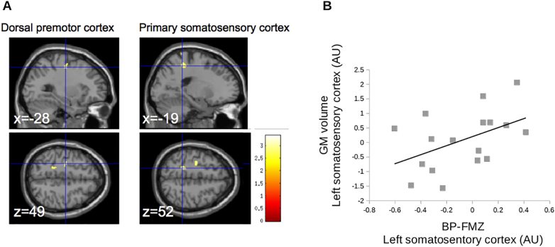
Results: In FHD patients compared to HV, the vermis VI of the right cerebellum and the left sensorimotor cortex had a decrease of Flumazenil binding potential (FMZ-BP), whereas the striatum and the lateral cerebellum did not show significant change. Bilateral inferior prefrontal cortex had increased FMZ-BP and an increase of perfusion, which correlated negatively with disease duration. Only the left sensorimotor cortex showed a decrease of grey matter volume.
Interpretation: Impairments of GABAergic neurotransmission in the cerebellum and the sensorimotor cortical areas could explain different aspects of loss of inhibitory control in FHD, the former being involved in maladaptive plasticity, the latter in surround inhibition. Reorganization of the inferior prefrontal cortices, part of the associative network, might be compensatory for the loss of inhibitory control in sensorimotor circuits. These findings suggest that cerebellar and cerebral GABAergic abnormalities could play a role in the functional imbalance of striato-cerebello-cortical loops in dystonia.
Keywords: Cerebellum; Focal dystonia; Inhibition; Motor cortex; Movement disorder.
Figures


Similar articles
-
In vivo evidence for GABA(A) receptor changes in the sensorimotor system in primary dystonia.Mov Disord. 2011 Apr;26(5):852-7. doi: 10.1002/mds.23553. Epub 2011 Mar 2. Mov Disord. 2011. PMID: 21370265
-
Individuated finger control in focal hand dystonia: an fMRI study.Neuroimage. 2012 Jul 16;61(4):823-31. doi: 10.1016/j.neuroimage.2012.03.066. Epub 2012 Mar 30. Neuroimage. 2012. PMID: 22484405 Free PMC article.
-
Network-specific resting-state connectivity changes in the premotor-parietal axis in writer's cramp.Neuroimage Clin. 2017 Oct 14;17:137-144. doi: 10.1016/j.nicl.2017.10.001. eCollection 2018. Neuroimage Clin. 2017. PMID: 29085775 Free PMC article.
-
Integrated technology for evaluation of brain function and neural plasticity.Phys Med Rehabil Clin N Am. 2004 Feb;15(1):263-306. doi: 10.1016/s1047-9651(03)00124-4. Phys Med Rehabil Clin N Am. 2004. PMID: 15029909 Review.
-
Hereditary dystonia as a neurodevelopmental circuit disorder: Evidence from neuroimaging.Neurobiol Dis. 2011 May;42(2):202-9. doi: 10.1016/j.nbd.2010.10.010. Epub 2010 Oct 19. Neurobiol Dis. 2011. PMID: 20965251 Free PMC article. Review.
Cited by
-
Cerebral blood flow in dystonia due to pantothenate kinase-associated neurodegeneration.Neuroradiol J. 2020 Dec;33(6):479-485. doi: 10.1177/1971400920943967. Epub 2020 Aug 27. Neuroradiol J. 2020. PMID: 32851917 Free PMC article.
-
Generalized dystonia unraveled: Molecular mechanisms, diagnostic strategies, and treatment paradigms.Neurol Sci. 2025 Aug 22. doi: 10.1007/s10072-025-08404-3. Online ahead of print. Neurol Sci. 2025. PMID: 40841848 Review.
-
Task-related brain activity and functional connectivity in upper limb dystonia: a functional magnetic resonance imaging (fMRI) and functional near-infrared spectroscopy (fNIRS) study.Neurophotonics. 2020 Oct;7(4):045004. doi: 10.1117/1.NPh.7.4.045004. Epub 2020 Oct 19. Neurophotonics. 2020. PMID: 33094125 Free PMC article.
-
Oscillatory Cortical Activity in an Animal Model of Dystonia Caused by Cerebellar Dysfunction.Front Cell Neurosci. 2018 Nov 6;12:390. doi: 10.3389/fncel.2018.00390. eCollection 2018. Front Cell Neurosci. 2018. PMID: 30459559 Free PMC article.
-
Neuroimaging Applications in Dystonia.Int Rev Neurobiol. 2018;143:1-30. doi: 10.1016/bs.irn.2018.09.007. Epub 2018 Oct 23. Int Rev Neurobiol. 2018. PMID: 30473192 Free PMC article. Review.
References
MeSH terms
Substances
Supplementary concepts
Grants and funding
LinkOut - more resources
Full Text Sources
Other Literature Sources
