

Structure-function relationship comparison between retinal nerve fibre layer and Bruch's membrane opening-minimum rim width in glaucoma
- PMID: 29062772
- PMCID: PMC5638974
- DOI: 10.18240/ijo.2017.10.09
Structure-function relationship comparison between retinal nerve fibre layer and Bruch's membrane opening-minimum rim width in glaucoma
Abstract
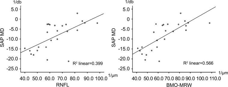
Aim: To evaluate and compare structural optical coherence tomography (OCT)-based parameters, such as Bruch's membrane opening-minimum rim width (BMO-MRW), and retinal nerve fiber layer (RNFL) thickness in glaucoma patients with visual field (VF) defects, and to correlate both to mean deviation (MD) values of obtained standard achromatic perimetry (SAP) examinations.
Methods: Patients with glaucoma and glaucomatous VF defects were enrolled in this prospective study and compared to age-matched healthy individuals. All study participants underwent a full ophthalmic examination and VF testing with SAP. Peripapillary RNFL thickness and BMO-MRW were acquired with SD-OCT. Correlation analyses between obtained global functional and global as well as sectorial structural parameters were calculated.
Results: A consecutive series of 30 glaucomatous right eyes of 30 patients were included and compared to 36 healthy right eyes of 36 individuals in the control group. Global MD of values correlated significantly with global RNFL (Pearson corr. coeff: 0.632, P=0.001) and global BMO-MRW (Pearson corr. coeff: 0.746, P<0.001) values in the glaucoma group. Global MD and sectorial RNFL or BMO-MRW values correlated less significantly. In the control group, MD values did not correlate with RNFL or BMO-MRW measurements. A subgroup analysis of myopic patients (>4 diopters) within the glaucoma group (n=6) revealed a tendency for higher correlations between MD and BMO-MRW than MD and RNFL measurements.
Conclusion: In a clinical setting, RNFL thickness and BMO-MRW correlate similarly with global VF sensitivity in glaucoma patients with BMO-MRW showing higher correlations in myopic glaucoma patients.
Keywords: Bruch's membrane opening-minimum rim width; glaucoma; myopia; optical coherence tomography; retinal nerve fibre layer; standard automated perimetry; visual field defects.
Figures
References
-
- Miglior S, Zeyen T, Pfeiffer N, Cunha-Vaz J, Torri V, Adamsons I, European Glaucoma Prevention Study (EGPS) Group Results of the European glaucoma prevention study. Ophthalmology. 2005;112(3):366–375. - PubMed
-
- Airaksinen PJ, Tuulonen A, Alanko HI. Rate and pattern of neuroretinal rim area decrease in ocular hypertension and glaucoma. Arch Ophthalmol. 1992;110(2):206–210. - PubMed
-
- Zangwill LM, Williams J, Berry CC, Knauer S, Weinreb RN. A comparison of optical coherence tomography and retinal nerve fiber layer photography for detection of nerve fiber layer damage in glaucoma. Ophthalmology. 2000;107(7):1309–1315. - PubMed
LinkOut - more resources
Full Text Sources
Other Literature Sources
Miscellaneous