. 2017 Oct 9:4:63.
doi: 10.3389/fcvm.2017.00063. eCollection 2017.

Toward Exosome-Based Therapeutics: Isolation, Heterogeneity, and Fit-for-Purpose Potency

Affiliations

Toward Exosome-Based Therapeutics: Isolation, Heterogeneity, and Fit-for-Purpose Potency

Gareth R Willis et al. Front Cardiovasc Med. .

Abstract

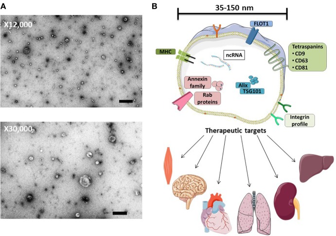
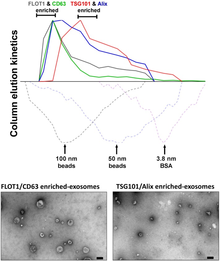
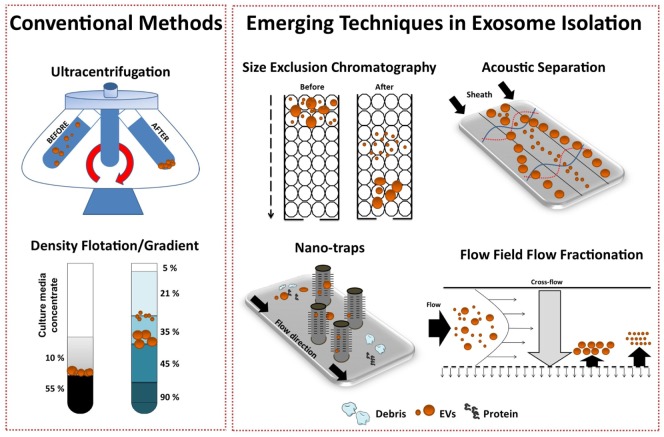
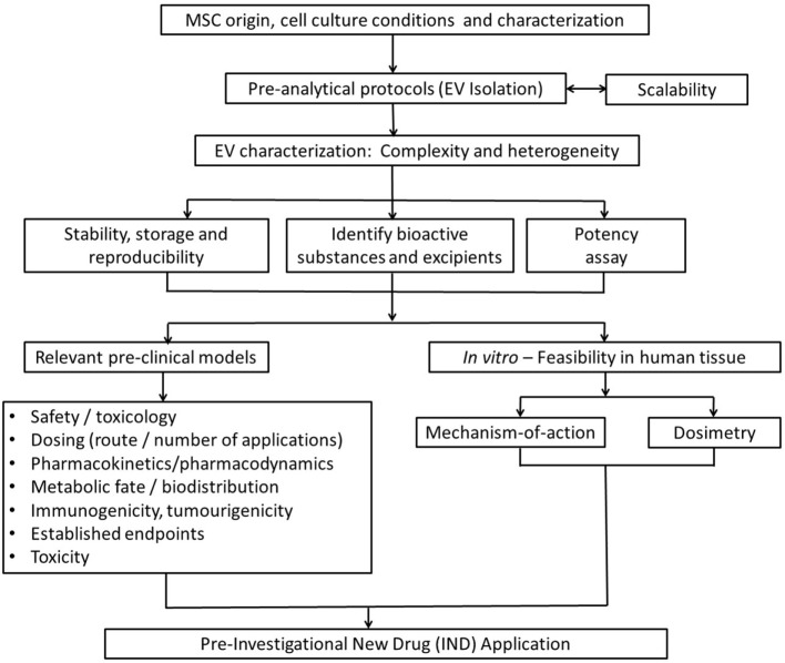
Exosomes are defined as submicron (30-150 nm), lipid bilayer-enclosed extracellular vesicles (EVs), specifically generated by the late endosomal compartment through fusion of multivesicular bodies with the plasma membrane. Produced by almost all cells, exosomes were originally considered to represent just a mechanism for jettisoning unwanted cellular moieties. Although this may be a major function in most cells, evolution has recruited the endosomal membrane-sorting pathway to duties beyond mere garbage disposal, one of the most notable examples being its cooption by retroviruses for the generation of Trojan virions. It is, therefore, tempting to speculate that certain cell types have evolved an exosome subclass active in intracellular communication. We term this EV subclass "signalosomes" and define them as exosomes that are produced by the "signaling" cells upon specific physiological or environmental cues and harbor cargo capable of modulating the programming of recipient cells. Our recent studies have established that signalosomes released by mesenchymal stem/stromal cells (MSCs) represent the main vector of MSC immunomodulation and therapeutic action in animal models of lung disease. The efficacy of MSC-exosome treatments in a number of preclinical models of cardiovascular and pulmonary disease supports the promise of application of exosome-based therapeutics across a wide range of pathologies within the near future. However, the full realization of exosome therapeutic potential has been hampered by the absence of standardization in EV isolation, and procedures for purification of signalosomes from the main exosome population. This is mainly due to immature methodologies for exosome isolation and characterization and our incomplete understanding of the specific characteristics and molecular composition of signalosomes. In addition, difficulties in defining metrics for potency of exosome preparations and the challenges of industrial scale-up and good manufacturing practice compliance have complicated smooth and timely transition to clinical development. In this manuscript, we focus on cell culture conditions, exosome harvesting, dosage, and exosome potency, providing some empirical guidance and perspectives on the challenges in bringing exosome-based therapies to clinic.

Keywords: exosome-based therapeutics; exosomes; extracellular vesicles; mesenchymal stem cells; preclinical.


Figures

Figure 1
Figure 1
MSC-exosome morphology and composition. (A) Transmission electron microscopy (TEM) images of human bone marrow-derived MSC-exosomes (low magnification, 12,000×, scale bar = 500 nm, and high magnification, 30,000×, scale bar = 100 nm) representative TEM images adapted from Ref. (8). (B) MSC-exosomes are surrounded by a phospholipid bilayer and may contain proteins, such as annexins (these are important for transport); tetraspanins such as CD9, CD81, and CD63; and other proteins, such as Alix and TSG101, that are involved in exosomal biogenesis from endosomes. MSC-exosome therapy has shown beneficial effects in numerous preclinical models, demonstrating histological and functional benefits in multiple organs. Abbreviations: FLOT1, flotillin-1; MHC, major histocompatibility complex; TSG101, tumor susceptibility gene 101.
Figure 2
Figure 2
Isolation of MSC-exosomes subtypes by size-exclusion chromatography (SEC). Briefly, exosomes were isolated directly from cell culture supernatants following a 36 h harvest period in serum-free-media. Cell culture media were subjected to differential centrifugation, 300 × g for 10 min, followed by 3,000 × g for 10 min, and 13,000 × g for 30 min to remove any cells, cell debris, and large apoptotic bodies in suspension, respectively. Conditioned media (CM) was concentrated 50-fold by tangential flow filtration (TFF) and the exosomes were purified using OptiPrep™ (iodixanol) cushion density flotation (3.5 h at 100,000 × g, 4°C), as previously described (8). Heterogeneous exosomes were further purified by size using size-exclusion chromatography (SEC). Here, sepharose CL-2B (80 mL) was washed with 1 × SSPE buffer [containing 1 mM EDTA and 149 mM NaCl in 0.20 mM phosphate buffer (pH 7.4)]. The column was packed with washed sepharose CL-2B to create a column with an internal diameter of 1.6 cm and height of 40 cm. Exosomes (1 ml, corresponding to 60 × 106 MSC equivalents) were added to column with a flow rate of 1 ml/min. Fractions (1 ml) were collected and assessed by dot plots and electron microscopy. The elution kinetics of 100 nm, 50 nm, and bovine serum albumin (BSA) were used to estimate exosome elution kinetics. TSG101, Alix, CD63, and FLOT1 levels were assessed by dot plots and are reported as relative intensity. Here, we identify two distinct MSC-exosome subtypes. The larger exosomes (>80 nm) have a greater flotillin-1 (FLOT1) and CD63 enrichment, while smaller exosomes (<80 nm) have a greater TSG101 and Alix ratio.
Figure 3
Figure 3
Conventional and emerging exosome isolation techniques. Ultracentrifugation (UC) is the most common exosome isolation method. Here, sedimentation of solutes including vesicles is governed by their size/density. Variations of UC such as layered or cushion based-density gradient UC are also widely employed. New methods are required to facilitate large-scale, high-yield production of exosomes for clinical applications. Several emerging technology platforms have shown promise in isolating exosomes from various sample matrices. Techniques, such as size-exclusion chromatography, ciliated micropillars nano-traps, acoustic wave separation technology, and flow field-flow fraction (F4), exploit unique biophysical traits of exosomes.
Figure 4
Figure 4
Stepwise approach to developing an MSC-exosome potency assay. Considering limitations in current exosome quantification techniques, assessment of exosome potency would be a valuable tool to standardize exosome dosing. Step 1: primary murine bone marrow-derived macrophages (BMDMs) were obtained by flushing the femur and tibia of 6- to 8-week-old FVB mice. M1 polarization was initiated by lipopolysaccharide (LPS) 100 ng/ml and interferon-γ (IFNγ) 20 ng/ml stimulation. Macrophage polarization (1 × 106 BMDMs) was initiated with/without the presence of MSC-exosome preparations (0.05–1 × 106 cell equivalents), providing several ratios of MSC-exosome cell equivalents-to-BMDMs. After 24 h, total RNA was isolated and TNFα mRNA levels were assessed by RT-qPCR. The functional endpoint is the capacity of MSC-exosomes to suppress the mRNA induction of TNFα. Step 2: an EC50 value (50% reduction in TNFα mRNA, relative to M1 control) is transformed to an arbitrary exosome potency unit (EPU). Data were adapted from our recent work (8), where we demonstrated purified human umbilical cord MSC-exosomes, dose dependently suppressed mRNA levels of TNFα in alveolar macrophages in vitro. Step 3: an EPU can be applied to standardize dosing between different exosome preparations or as a means of correlating potency to exosome quantity.
Figure 5
Figure 5
Strategic flowchart for the preclinical testing of exosome-based therapeutics. EV, extracellular vesicle. Adapted from Ref. (97).

References

    1. Gould SJ, Raposo G. As we wait: coping with an imperfect nomenclature for extracellular vesicles. J Extracell Vesicles (2013) 2:20389. 10.3402/jev.v2i0.20389 - DOI - PMC - PubMed
    1. Willms E, Johansson HJ, Mäger I, Lee Y, Blomberg KEM, Sadik M, et al. Cells release subpopulations of exosomes with distinct molecular and biological properties. Sci Rep (2016) 6:22519. 10.1038/srep22519 - DOI - PMC - PubMed
    1. Johnstone RM, Adam M, Hammond JR, Orr L, Turbide C. Vesicle formation during reticulocyte maturation. Association of plasma membrane activities with released vesicles (exosomes). J Biol Chem (1987) 262:9412–20. - PubMed
    1. Théry C. Exosomes: secreted vesicles and intercellular communications. F1000 Biol Rep (2011) 3:15. 10.3410/B3-15 - DOI - PMC - PubMed
    1. van der Pol E, Böing AN, Harrison P, Sturk A, Nieuwland R. Classification, functions, and clinical relevance of extracellular vesicles. Pharmacol Rev (2012) 64:676–705. 10.1124/pr.112.005983 - DOI - PubMed