Review
. 2017 Oct 23;5(1):49.
doi: 10.1186/s40635-017-0163-0.

Assessment of immune organ dysfunction in critical illness: utility of innate immune response markers

Affiliations
Review

Assessment of immune organ dysfunction in critical illness: utility of innate immune response markers

Carmen Andrea Pfortmueller et al. Intensive Care Med Exp. .

Abstract

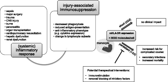
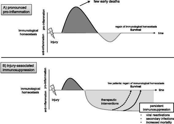
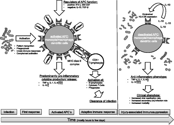
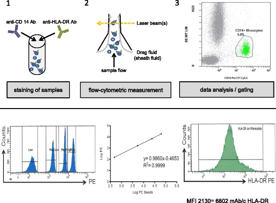
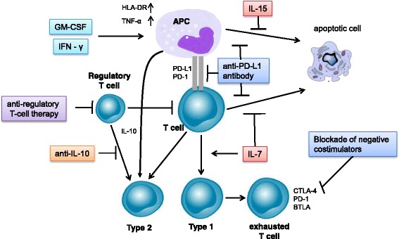
In critically ill patients, organ dysfunctions are routinely assessed, monitored, and treated. Mounting data show that substantial critical illness-induced changes in the immune system can be observed in most ICU patients and that not only "hyper-inflammation" but also persistence of an anti-inflammatory phenotype (as in sepsis-associated immunosuppression) is associated with increased morbidity and mortality. Despite common perception, changes in functional immunity cannot be adequately assessed by routine inflammatory biomarkers such as C-reactive protein, procalcitonin, or numerical analysis of leukocyte (sub)-counts. Cytokines appear also not suited due to their short half-life and pleiotropy, their unexclusive origin from immune cells, and their potential to undergo antagonization by circulating inactivating molecules. Thus, beyond leukocyte quantification and use of routine biomarkers, direct assessment of immune cell function seems required to characterize the immune systems' status. This may include determination of, e.g., ex vivo cellular cytokine release, phagocytosis activity, and/or antigen-presenting capacity. In this regard, standardized flow-cytometric assessment of the major histocompatibility-II complex human leukocyte antigen (-D related) (HLA-DR) has gained particular interest. Monocytic HLA-DR (mHLA-DR) controls the interplay between innate and adaptive immunity and may serve as a "global" biomarker of injury-associated immunosuppression, and its decreased expression is associated with adverse clinical outcomes (e.g., secondary infection risk, mortality). Importantly, recent data demonstrate that injury-associated immunosuppression can be reversed-opening up new therapeutic avenues in affected patients. Here we discuss the potential scientific and clinical value of assessment of functional immunity with a focus on monocytes/macrophages and review the current state of knowledge and potential perspectives for affected critically ill patients.

Keywords: Biomarkers; Critical illness; HLA-DR expression; Immune function; Immune modulation; Immune suppression; Immunomodulation; Sepsis; Sepsis-associated immunosuppression; mHLA-DR.


Conflict of interest statement

Ethics approval and consent to participate

Not applicable.

Competing interests

The authors declare that they have no competing interests.

Publisher’s Note

Springer Nature remains neutral with regard to jurisdictional claims in published maps and institutional affiliations.

Figures

Fig. 1
Fig. 1
Injury-associated immunosuppression in critically ill patients. Injury-associated immunosuppression (IAI) may develop in critical illness. IAI was shown to be of importance in cases of persistence for ≥ 2 days. Key future potential therapeutical options are listed. Monocytic HLA-DR expression (mHLA-DR, given in bound antibodies per cell) may serve as a global marker of IAI
Fig. 2
Fig. 2
Inter-individual injury-associated response patterns in critically ill patients. Patients with critical illness respond differently to injury (e.g., sepsis). Whereas patient “A” undergoes a pronounced inflammatory phase (net effects are shown) with regain of immunological homeostasis and subsequent survival, patient “B” enters a persisting phase of injury-associated immunosuppression (IAI). In IAI, viral reactivation rates, secondary (re-) infection rates, and mortality is increased. This underlines the importance of inter-individual response patterns and need for individual patient characterization before application of interventional therapeutic approaches (adapted from Hotchkiss et al., 2013 [4])
Fig. 3
Fig. 3
Infection-induced activation of key immune cells. In sepsis, bacterial infections trigger numerous pathways resulting in activation of key antigen-presenting cells (APCs) (i.e., monocytes/macrophages, dendritic cells). Activated APCs predominantly express pro-inflammatory cytokines and present antigens bound to major histocompatibility (MHC) class II complexes (such as HLA-DR). Antigen-bound HLA-DR triggers T-cell-receptor (TCR) and co-stimulatory molecule (e.g. CD 40-CD40L) binding. Adaptive immune responses are initiated resulting in clearance of infection. In, e.g., cases of overwhelming infection, deactivation of monocytes, as in sepsis-associated immunosuppression (SAI), may occur. SAI is characterized by a shift towards an anti-inflammatory phenotype with predominant expression of IL-10 and diminished HLA-DR expression, resulting in impaired clearance of infection and increased mortality. In IAI, the deactivated phenotype can be observed immediately after injury
Fig. 4
Fig. 4
Flow-cytometric assessment of monocytic HLA-DR expression. Upper row: after staining of EDTA samples with specific antibodies, HLA-DR expression is assessed on CD14+ monocytes by flow cytometry. Lower row: (left) Quantibrite™-PE beads are used to calculate a calibration curve (middle) for HLA-DR assessment on CD14+ monocytes. (Right) mean fluorescence intensity (MFI) values for HLA-DR on monocytes are converted in a given sample to molecules per cell using the calibration curve
Fig. 5
Fig. 5
Potential future immunomodulatory approaches in sepsis. Key approaches to reverse sepsis-associated immunosuppression include cytokine-induced stimulation of monocyte/macrophage function (GM-CSF, IFN-γ), administration of survival factors for T cells (IL-7), blockade of anti-inflammatory mechanisms (anti-IL-10 antibody/antagonization of regulatory T-cell function), approaches to target immune cell exhaustion/apoptosis (anti-programmed death (PD) receptor 1 or PD-ligand1 (PD-L1)), and blockade of negative co-stimulators (e.g., cytotoxic T-lymphocyte-associated protein 4 [CTLA-4] or B- and T-lymphocyte attenuator (BTLA))

References

    1. Rieckmann JC, Geiger R, Hornburg D, Wolf T, Kveler K, Jarrossay D, Sallusto F, Shen-Orr SS, Lanzavecchia A, Mann M, et al. Social network architecture of human immune cells unveiled by quantitative proteomics. Nat Immunol. 2017;18(5):583–593. doi: 10.1038/ni.3693. - DOI - PubMed
    1. Schefold JC, Hasper D, Reinke P, Monneret G, Volk HD. Consider delayed immunosuppression into the concept of sepsis. Crit Care Med. 2008;36(11):3118. doi: 10.1097/CCM.0b013e31818bdd8f. - DOI - PubMed
    1. Carlet J, Cohen J, Calandra T, Opal SM, Masur H. Sepsis: time to reconsider the concept. Crit Care Med. 2008;36(3):964–966. doi: 10.1097/CCM.0B013E318165B886. - DOI - PubMed
    1. Hotchkiss RS, Monneret G, Payen D. Immunosuppression in sepsis: a novel understanding of the disorder and a new therapeutic approach. Lancet Infect Dis. 2013;13(3):260–268. doi: 10.1016/S1473-3099(13)70001-X. - DOI - PMC - PubMed
    1. Schefold JC, Hasper D, Volk HD, Reinke P. Sepsis: time has come to focus on the later stages. Med Hypotheses. 2008;71(2):203–208. doi: 10.1016/j.mehy.2008.03.022. - DOI - PubMed