

Non-invasive imaging of global and regional cardiac function in pulmonary hypertension
- PMID: 29064323
- PMCID: PMC5753990
- DOI: 10.1177/2045893217742000
Non-invasive imaging of global and regional cardiac function in pulmonary hypertension
Abstract
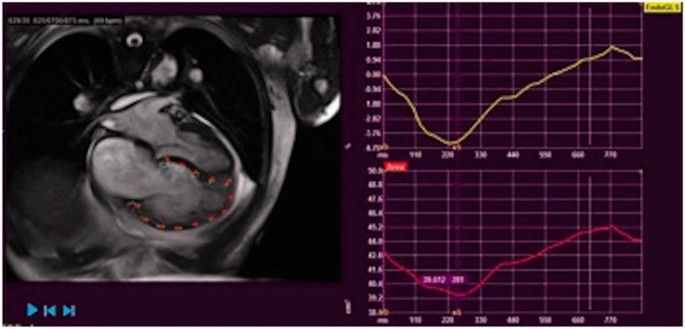
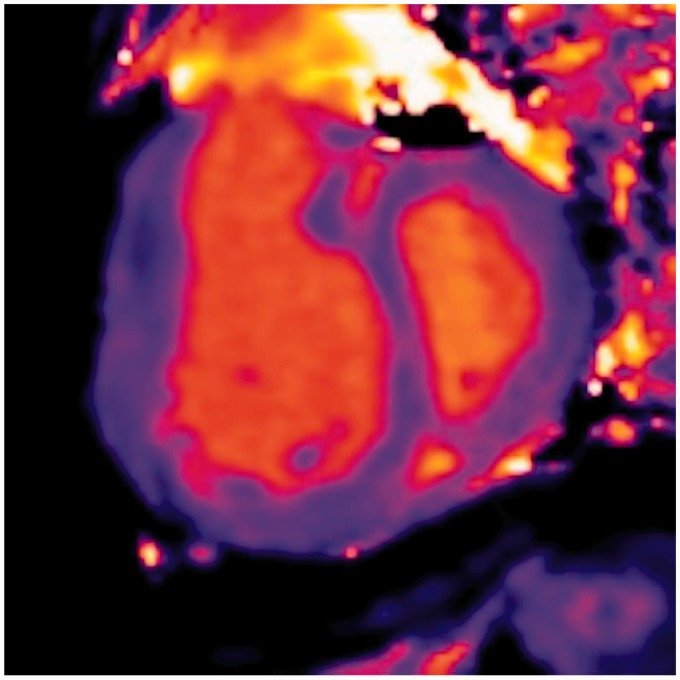
Pulmonary hypertension (PH) is a progressive illness characterized by elevated pulmonary artery pressure; however, the main cause of mortality in PH patients is right ventricular (RV) failure. Historically, improving the hemodynamics of pulmonary circulation was the focus of treatment; however, it is now evident that cardiac response to a given level of pulmonary hemodynamic overload is variable but plays an important role in the subsequent prognosis. Non-invasive tests of RV function to determine prognosis and response to treatment in patients with PH is essential. Although the right ventricle is the focus of attention, it is clear that cardiac interaction can cause left ventricular dysfunction, thus biventricular assessment is paramount. There is also focus on the atrial chambers in their contribution to cardiac function in PH. Furthermore, there is evidence of regional dysfunction of the two ventricles in PH, so it would be useful to understand both global and regional components of dysfunction. In order to understand global and regional cardiac function in PH, the most obvious non-invasive imaging techniques are echocardiography and cardiac magnetic resonance imaging (CMRI). Both techniques have their advantages and disadvantages. Echocardiography is widely available, relatively inexpensive, provides information regarding RV function, and can be used to estimate RV pressures. CMRI, although expensive and less accessible, is the gold standard of biventricular functional measurements. The advent of 3D echocardiography and techniques including strain analysis and stress echocardiography have improved the usefulness of echocardiography while new CMRI technology allows the measurement of strain and measuring cardiac function during stress including exercise. In this review, we have analyzed the advantages and disadvantages of the two techniques and discuss pre-existing and novel forms of analysis where echocardiography and CMRI can be used to examine atrial, ventricular, and interventricular function in patients with PH at rest and under stress.
Keywords: echocardiography; magnetic resonance imaging; pulmonary arterial hypertension; pulmonary hypertension; right ventricle function and dysfunction.
Figures
References
-
- D’Alto M, Scognamiglio G, Dimopoulos K, et al. Right heart and pulmonary vessels structure and function. Echocardiography 2015; 32(Suppl. 1): S3–10. - PubMed
-
- Guazzi M, Naeije R, Arena R, et al. Echocardiography of right ventriculoarterial coupling combined with cardiopulmonary exercise testing to predict outcome in heart failure. Chest 2015; 148(1): 226–234. - PubMed
LinkOut - more resources
Full Text Sources
Other Literature Sources
