

Dermaseptin-PH: A Novel Peptide with Antimicrobial and Anticancer Activities from the Skin Secretion of the South American Orange-Legged Leaf Frog, Pithecopus (Phyllomedusa) hypochondrialis
- PMID: 29064402
- PMCID: PMC6151546
- DOI: 10.3390/molecules22101805
Dermaseptin-PH: A Novel Peptide with Antimicrobial and Anticancer Activities from the Skin Secretion of the South American Orange-Legged Leaf Frog, Pithecopus (Phyllomedusa) hypochondrialis
Abstract
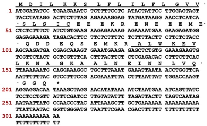
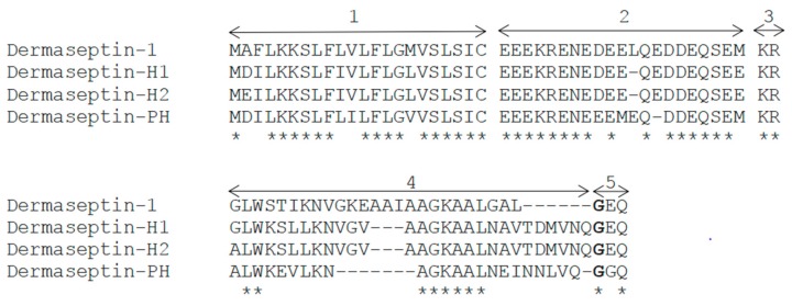
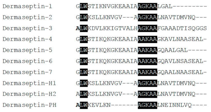
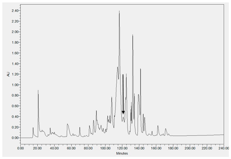
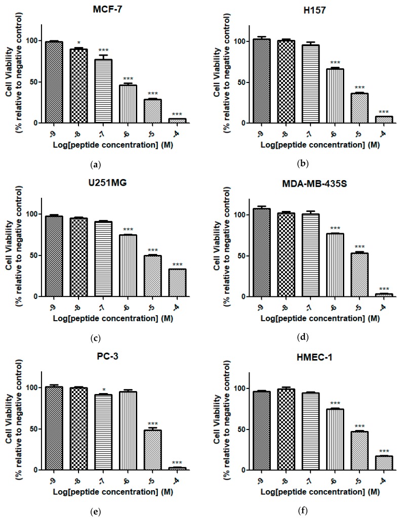
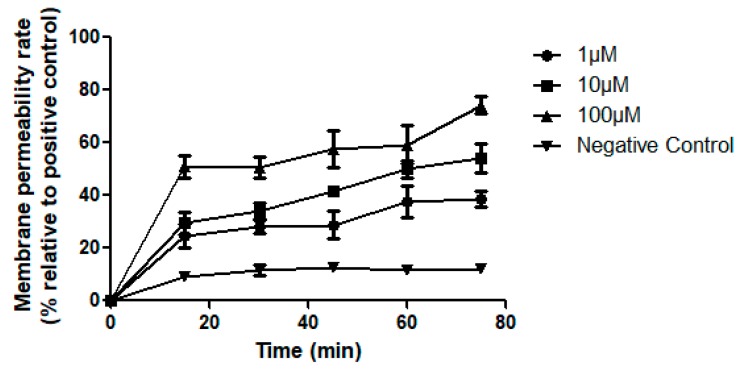
The dermaseptin peptides, mainly derived from the skin secretions of Hylidae frogs, belong to a superfamily of antimicrobial peptides and exhibit diverse antimicrobial and anticancer activities with low cytotoxicity. Here, we reported a novel dermaseptin peptide, from the South American orange-legged leaf frogs, Pithecopus (Phyllomedusa) hypochondrialis, processing the shortest peptide length, namely Dermaseptin-PH. The complementary DNA (cDNA) encoding biosynthetic precursor of Dermaseptin-PH was initially identified by the rapid amplification of cDNA ends PCR (RACE-PCR) technique from the skin secretion. The predicted primary structure was confirmed by a combination of reverse-phase high performance liquid chromatography (RP-HPLC) and MS/MS fragmentation from the skin secretion. Chemically-synthetic Dermaseptin-PH was investigated using a range of bioactivity assessment assays to evaluate the biological activities and cytotoxicity of Dermaseptin-PH. Dermaseptin-PH inhibited the growth of Gram-negative bacteria, Gram-positive bacteria, and pathogenic yeast Candidaalbicans. In addition, Dermaseptin-PH showed a broad-spectrum of anticancer activities against several cancer cell lines including MCF-7, H157, U251MG, MDA-MB-435S, and PC-3. The potent antimicrobial and anticancer activities of Dermaseptin-PH make it a promising candidate in the discovery of new drugs for clinical applications, and the relatively short sequence of Dermaseptin-PH can provide new insight for the research and structural modification of new peptide drugs.
Keywords: amphibian skin secretion; anticancer; antimicrobial; dermaseptin; molecular cloning.
Conflict of interest statement
The authors declare no conflict of interest.
Figures
Similar articles
-
Two Novel Dermaseptin-Like Antimicrobial Peptides with Anticancer Activities from the Skin Secretion of Pachymedusa dacnicolor.Toxins (Basel). 2016 May 12;8(5):144. doi: 10.3390/toxins8050144. Toxins (Basel). 2016. PMID: 27187467 Free PMC article.
-
Evaluating the Bioactivity of a Novel Antimicrobial and Anticancer Peptide, Dermaseptin-PS4(Der-PS4), from the Skin Secretion of Phyllomedusa sauvagii.Molecules. 2019 Aug 16;24(16):2974. doi: 10.3390/molecules24162974. Molecules. 2019. PMID: 31426323 Free PMC article.
-
Biological Activities of Cationicity-Enhanced and Hydrophobicity-Optimized Analogues of an Antimicrobial Peptide, Dermaseptin-PS3, from the Skin Secretion of Phyllomedusa sauvagii.Toxins (Basel). 2018 Aug 7;10(8):320. doi: 10.3390/toxins10080320. Toxins (Basel). 2018. PMID: 30087268 Free PMC article.
-
The dermaseptin superfamily: a gene-based combinatorial library of antimicrobial peptides.Biochim Biophys Acta. 2009 Aug;1788(8):1537-50. doi: 10.1016/j.bbamem.2008.09.006. Epub 2008 Sep 26. Biochim Biophys Acta. 2009. PMID: 18929530 Review.
-
Dermaseptins as models for the elucidation of membrane-acting helical amphipathic antimicrobial peptides.Curr Pharm Biotechnol. 2011 Aug;12(8):1184-93. doi: 10.2174/138920111796117319. Curr Pharm Biotechnol. 2011. PMID: 21470155 Review.
Cited by
-
Exploration of the Structure-Function Relationships of a Novel Frog Skin Secretion-Derived Bioactive Peptide, t-DPH1, through Use of Rational Design, Cationicity Enhancement and In Vitro Studies.Antibiotics (Basel). 2021 Dec 14;10(12):1529. doi: 10.3390/antibiotics10121529. Antibiotics (Basel). 2021. PMID: 34943741 Free PMC article.
-
Biofilms: Novel Strategies Based on Antimicrobial Peptides.Pharmaceutics. 2019 Jul 10;11(7):322. doi: 10.3390/pharmaceutics11070322. Pharmaceutics. 2019. PMID: 31295834 Free PMC article. Review.
-
A Novel Amphibian Antimicrobial Peptide, Phylloseptin-PV1, Exhibits Effective Anti-staphylococcal Activity Without Inducing Either Hepatic or Renal Toxicity in Mice.Front Microbiol. 2020 Oct 26;11:565158. doi: 10.3389/fmicb.2020.565158. eCollection 2020. Front Microbiol. 2020. PMID: 33193152 Free PMC article.
-
Biodegradable Polymers and Polymer Composites with Antibacterial Properties.Int J Mol Sci. 2023 Apr 18;24(8):7473. doi: 10.3390/ijms24087473. Int J Mol Sci. 2023. PMID: 37108637 Free PMC article. Review.
-
A phylogenetic review of cancer resistance highlights evolutionary solutions to Peto's Paradox.Genet Mol Biol. 2022 Dec 5;45(3 Suppl 1):e20220133. doi: 10.1590/1678-4685-GMB-2022-0133. eCollection 2022. Genet Mol Biol. 2022. PMID: 36534348 Free PMC article.
References
-
- Da Cunha N.B., Cobacho N.B., Viana J.F., Lima L.A., Sampaio K.B., Dohms S.S., Ferreira A.C., de la Fuente-Nunez C., Costa F.F., Franco O.L., et al. The next generation of antimicrobial peptides (AMPs) as molecular therapeutic tools for the treatment of diseases with social and economic impacts. Drug Discov. Today. 2017;22:234–248. doi: 10.1016/j.drudis.2016.10.017. - DOI - PMC - PubMed
MeSH terms
Substances
LinkOut - more resources
Full Text Sources
Other Literature Sources
Molecular Biology Databases
Miscellaneous
