Review
. 2017 Oct 24;7(10):138.
doi: 10.3390/brainsci7100138.

Pattern Recognition of the Multiple Sclerosis Syndrome

Affiliations
Review

Pattern Recognition of the Multiple Sclerosis Syndrome

Rana K Zabad et al. Brain Sci. .

Abstract

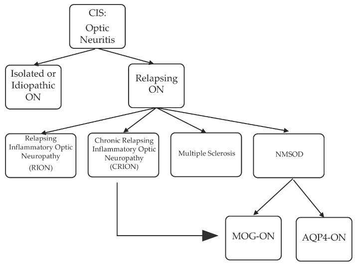
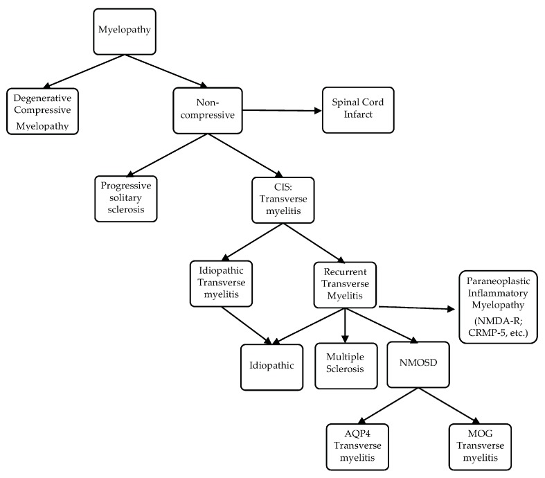
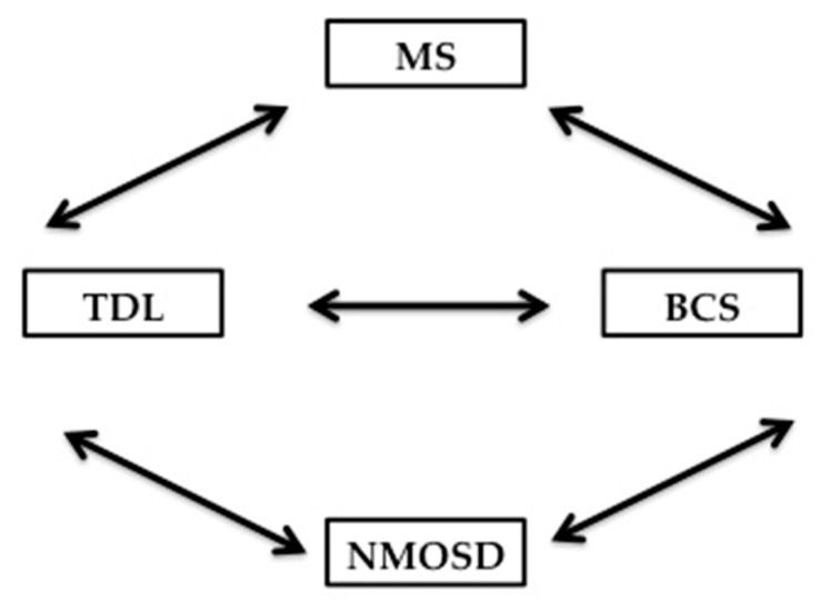
During recent decades, the autoimmune disease neuromyelitis optica spectrum disorder (NMOSD), once broadly classified under the umbrella of multiple sclerosis (MS), has been extended to include autoimmune inflammatory conditions of the central nervous system (CNS), which are now diagnosable with serum serological tests. These antibody-mediated inflammatory diseases of the CNS share a clinical presentation to MS. A number of practical learning points emerge in this review, which is geared toward the pattern recognition of optic neuritis, transverse myelitis, brainstem/cerebellar and hemispheric tumefactive demyelinating lesion (TDL)-associated MS, aquaporin-4-antibody and myelin oligodendrocyte glycoprotein (MOG)-antibody NMOSD, overlap syndrome, and some yet-to-be-defined/classified demyelinating disease, all unspecifically labeled under MSsyndrome. The goal of this review is to increase clinicians' awareness of the clinical nuances of the autoimmune conditions for MS and NMSOD, and to highlight highly suggestive patterns of clinical, paraclinical or imaging presentations in order to improve differentiation. With overlay in clinical manifestations between MS and NMOSD, magnetic resonance imaging (MRI) of the brain, orbits and spinal cord, serology, and most importantly, high index of suspicion based on pattern recognition, will help lead to the final diagnosis.

Keywords: AQP4 antibodies; MOG antibodies; MS; NMOSD; brainstem syndrome; clinically isolated syndrome (CIS); optic neuritis; transverse myelitis; tumefactive demyelinating lesions.


Conflict of interest statement

Rana K. Zabad has been a site investigator or site PI for clinical trials funded by Biogen, Genentech, Novartis, Sunpharma. In the last 2 years, she has served as a consultant for Bayer, Genzyme, TEVA Neuroscience and TG therapeutics and has given unbranded lectures sponsored by TEVA and is a member of the Adjudication Committee for a clinical trial of biotin in primary and secondary progressive multiple sclerosis sponsored by PAREXEL and medDay pharmaceutical. Renee Stewart has nothing to disclose. Kathleen Healey has received funding in the past for investigation-initiated research from the Multiple Sclerosis Foundation. In relation to this review specifically, the authors declare no conflict of interest.

Figures

Figure 1
Figure 1
Evolution of clinically isolated syndrome (CIS) optic neuritis: The majority of patients with ON will evolve into RION, CRION, MS or NMOSD. A percentage of patients will stay as isolated ON. Furthermore, there is more data that at least a subset of patients with CRION are indeed MOG-antibody associated NMOSD. AQP4-ON: Aquaporin 4-antibody-associated optic neuritis; MOG-ON: Myelin oligodendrocyte glycoprotein-antibody-associated optic neuritis.
Figure 2
Figure 2
Axial short tau inversion recovery (STIR and T1 with contrast orbital MRI of an 18-year-old Caucasian male, with bilateral longitudinally extensive (small arrows) optic neuritis with anterior predominance, perineural sheath swelling (long arrows) and tilting and twisting of both optic nerves better seen on axial STIR (2a,2b). Serum MOG-antibody was positive.
Figure 3
Figure 3
Evolution of clinically isolated syndrome (CIS) transverse myelitis.
Figure 4
Figure 4
55-year-old African-American female, with AQP4-NMOSD; sagittal STIR cervical MRI demonstrates simultaneous linear lesion and LETM from the medulla to C4 (4a) seen at the center of the cord on axial T2 (4b).
Figure 5
Figure 5
Added to above paragraph 55-year-old African-American female, with AQP4-NMOSD sagittal STIR cervical MRI demonstrates simultaneous linear lesion and LETM from the medulla to C4 (5a). An axial cut at the level of the cervicomedullary junction, axial T2 demonstrates 4 peripheral bright spotty lesions (5b).
Figure 6
Figure 6
Diagram summarizing the relationship between TDL, BCS, MS and NMOSD; TDL: Tumefactive demyelinating lesion; BCS: Balo’s concentric sclerosis; MS: Multiple sclerosis; NMOSD: Neuromyelitis optica spectrum disorder.
Figure 7
Figure 7
This is the case of a 21-year-old woman who presented with a history of bilateral optic neuritis at the age of 10. She subsequently had recurrent encephalitis with cortical swelling and seizures at the age of 13, 17 and 19. Axial FLAIR shows a right parietal cortical T2 hyperintensity with swelling (7a) associated with minimal T1 hypointensity without contrast enhancement (7b). Recurrent encephalitis was associated with a new right frontal cortical T2 hyperintensity with swelling (7c) associated with minimal T1 hypointensity with contrast enhancement. (7d). A third episode demonstrated a new left frontal cortical T2 hyperintensity with swelling (7e) associated with minimal T1 hypointensity with contrast enhancement (7f). Serum MOG-antibody was positive.

Similar articles

Cited by

References

    1. Fang B., McKeon A., Hinson S.R., Kryzer T.J., Pittock S.J., Aksamit A.J., Lennon V.A. Autoimmune Glial Fibrillary Acidic Protein Astrocytopathy: A Novel Meningoencephalomyelitis. JAMA Neurol. 2016;73:1297–1307. doi: 10.1001/jamaneurol.2016.2549. - DOI - PubMed
    1. Holtje M., Mertens R., Schou M.B., Saether S.G., Kochova E., Jarius S., Pruss H., Komorowski L., Probst C., Paul F., et al. Synapsin-antibodies in psychiatric and neurological disorders: Prevalence and clinical findings. Brain Behav. Immun. 2017;66:125–134. doi: 10.1016/j.bbi.2017.07.011. - DOI - PubMed
    1. Zamvil S.S., Slavin A.J. Does MOG Ig-positive AQP4-seronegative opticospinal inflammatory disease justify a diagnosis of NMO spectrum disorder? Neurol. Neuroimmunol. Neuroinflamm. 2015;2:e62. doi: 10.1212/NXI.0000000000000062. - DOI - PMC - PubMed
    1. Zekeridou A., Lennon V.A. Aquaporin-4 autoimmunity. Neurol. Neuroimmunol. Neuroinflamm. 2015;2:e110. doi: 10.1212/NXI.0000000000000110. - DOI - PMC - PubMed
    1. Poser C.M., Brinar V.V. Diagnostic criteria for multiple sclerosis: An historical review. Clin. Neurol. Neurosurg. 2004;106:147–158. doi: 10.1016/j.clineuro.2004.02.004. - DOI - PubMed

LinkOut - more resources