

Senescence-associated ultrastructural features of long-term cultures of induced pluripotent stem cells (iPSCs)
- PMID: 29064821
- PMCID: PMC5680563
- DOI: 10.18632/aging.101309
Senescence-associated ultrastructural features of long-term cultures of induced pluripotent stem cells (iPSCs)
Abstract
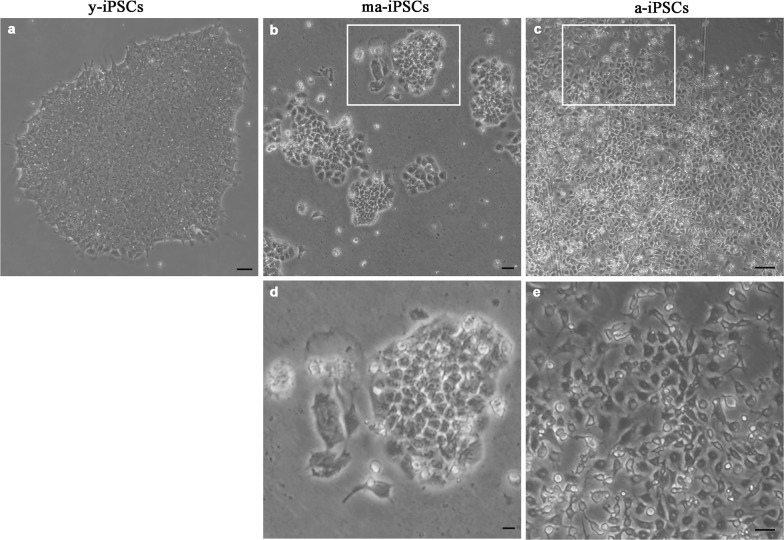
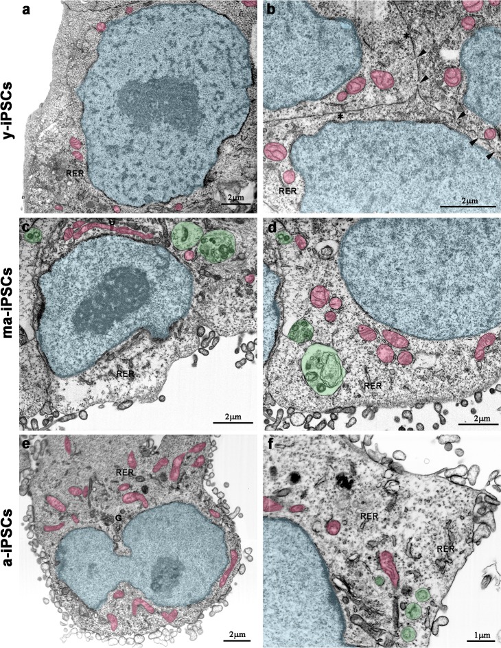
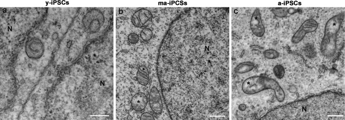
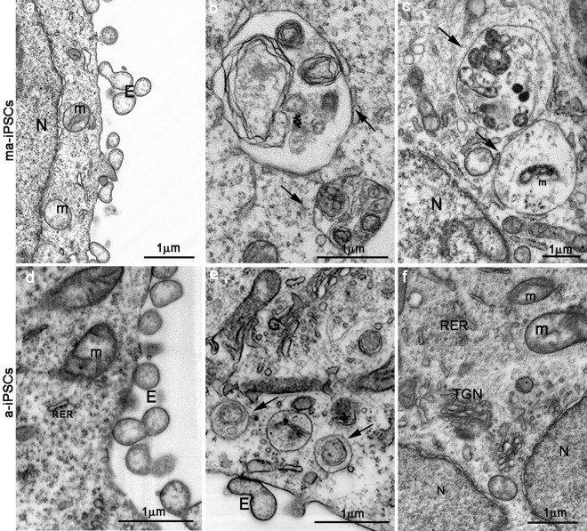
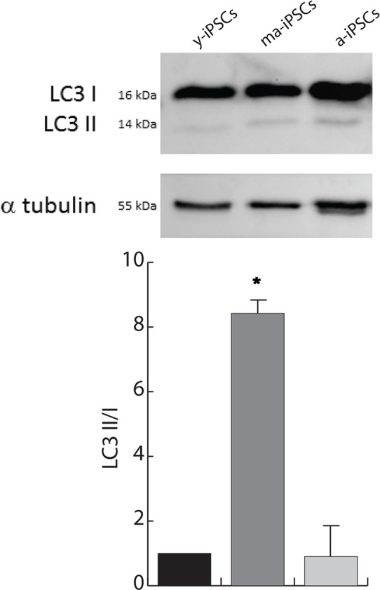
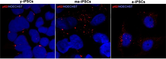
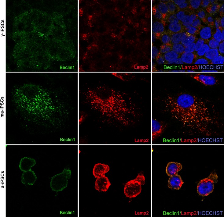
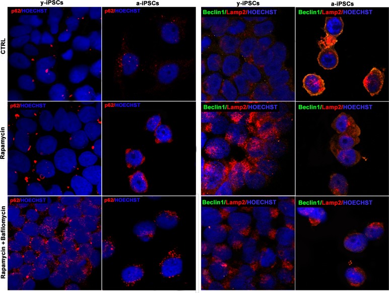
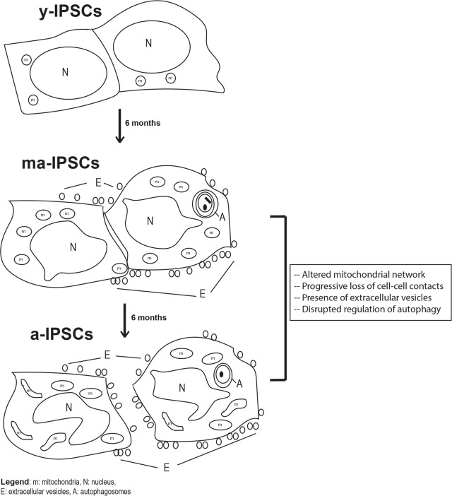
Induced pluripotent stem cells (iPSCs) hold great promise for developing personalized regenerative medicine, however characterization of their biological features is still incomplete. Moreover, changes occurring in long-term cultured iPSCs have been reported, suggesting these as a model of cellular aging. For this reason, we addressed the ultrastructural characterization of iPSCs, with a focus on possible time-dependent changes, involving specific cell compartments. To this aim, we comparatively analysed cultures at different timepoints, by an innovative electron microscopic technology (FIB/SEM). We observed progressive loss of cell-to-cell contacts, associated with increased occurrence of exosomes. Mitochondria gradually increased, while acquiring an elongated shape, with well-developed cristae. Such mitochondrial maturation was accompanied by their turnover, as assessed by the presence of autophagomes (undetectable in young iPSCs), some containing recognizable mitochondria. This finding was especially frequent in middle-aged iPSCs, while being occasional in aged cells, suggesting early autophagic activation followed by a decreased efficiency of the process with culturing time. Accordingly, confocal microscopy showed age-dependent alterations to the expression and distribution of autophagic markers. Interestingly, responsivity to rapamycin, highest in young iPSCs, was almost lost in aged cells. Overall, our results strongly support long-term cultured iPSCs as a model for studying relevant aspects of cellular senescence, involving intercellular communication, energy metabolism, and autophagy.
Keywords: FIB/SEM; aging; autophagy; cell-cell contacts; iPSCs; mitochondria.
Conflict of interest statement
The authors of this manuscript declare no conflict of interests.
Figures
Similar articles
-
The senescence-related mitochondrial/oxidative stress pathway is repressed in human induced pluripotent stem cells.Stem Cells. 2010 Apr;28(4):721-33. doi: 10.1002/stem.404. Stem Cells. 2010. PMID: 20201066
-
Exosomes Derived from Human Induced Pluripotent Stem Cells Ameliorate the Aging of Skin Fibroblasts.Int J Mol Sci. 2018 Jun 9;19(6):1715. doi: 10.3390/ijms19061715. Int J Mol Sci. 2018. PMID: 29890746 Free PMC article.
-
Aged induced pluripotent stem cell (iPSCs) as a new cellular model for studying premature aging.Aging (Albany NY). 2017 May 31;9(5):1453-1469. doi: 10.18632/aging.101248. Aging (Albany NY). 2017. PMID: 28562315 Free PMC article.
-
The Potential of iPSCs for the Treatment of Premature Aging Disorders.Int J Mol Sci. 2017 Nov 7;18(11):2350. doi: 10.3390/ijms18112350. Int J Mol Sci. 2017. PMID: 29112121 Free PMC article. Review.
-
Mitochondrial function in pluripotent stem cells and cellular reprogramming.Gerontology. 2014;60(2):174-82. doi: 10.1159/000355050. Epub 2013 Nov 19. Gerontology. 2014. PMID: 24281332 Review.
Cited by
-
Mitochondrial and Peroxisomal Alterations Contribute to Energy Dysmetabolism in Riboflavin Transporter Deficiency.Oxid Med Cell Longev. 2020 Aug 12;2020:6821247. doi: 10.1155/2020/6821247. eCollection 2020. Oxid Med Cell Longev. 2020. PMID: 32855765 Free PMC article.
-
Guidelines for the use and interpretation of assays for monitoring autophagy (4th edition)1.Autophagy. 2021 Jan;17(1):1-382. doi: 10.1080/15548627.2020.1797280. Epub 2021 Feb 8. Autophagy. 2021. PMID: 33634751 Free PMC article.
-
Clinical Potential of Cellular Material Sources in the Generation of iPSC-Based Products for the Regeneration of Articular Cartilage.Int J Mol Sci. 2023 Sep 22;24(19):14408. doi: 10.3390/ijms241914408. Int J Mol Sci. 2023. PMID: 37833856 Free PMC article. Review.
-
Induction of functional neutrophils from mouse fibroblasts by thymidine through enhancement of Tet3 activity.Cell Mol Immunol. 2022 May;19(5):619-633. doi: 10.1038/s41423-022-00842-9. Epub 2022 Mar 17. Cell Mol Immunol. 2022. PMID: 35301470 Free PMC article.
-
Mitochondrial dysfunction in mandibular hypoplasia, deafness and progeroid features with concomitant lipodystrophy (MDPL) patients.Aging (Albany NY). 2022 Feb 23;14(4):1651-1664. doi: 10.18632/aging.203910. Epub 2022 Feb 23. Aging (Albany NY). 2022. PMID: 35196257 Free PMC article.
References
-
- Takahashi K, Yamanaka S. Induction of pluripotent stem cells from mouse embryonic and adult fibroblast cultures by defined factors. Cell. 2006;126:663–76. https://doi.org/10.1016/j.cell.2006.07.024 - DOI - PubMed
-
- Compagnucci C, Nizzardo M, Corti S, Zanni G, Bertini E. In vitro neurogenesis: development and functional implications of iPSC technology. Cell Mol Life Sci. 2014;71:1623–39. https://doi.org/10.1007/s00018-013-1511-1 - DOI - PMC - PubMed
-
- Bukowiecki R, Adjaye J, Prigione A. Mitochondrial function in pluripotent stem cells and cellular reprogramming. Gerontology. 2014;60:174–82. https://doi.org/10.1159/000355050 - DOI - PubMed
-
- Raikwar SP, Kim EM, Sivitz WI, Allamargot C, Thedens DR, Zavazava N. Human iPS cell-derived insulin producing cells form vascularized organoids under the kidney capsules of diabetic mice. PLoS One. 2015;10:e0116582. https://doi.org/10.1371/journal.pone.0116582 - DOI - PMC - PubMed
-
- Beckmann A, Schubert M, Hainz N, Haase A, Martin U, Tschernig T, Meier C. Ultrastructural demonstration of Cx43 gap junctions in induced pluripotent stem cells from human cord blood. Histochem Cell Biol. 2016;146:529–37. https://doi.org/10.1007/s00418-016-1469-9 - DOI - PubMed
Publication types
MeSH terms
LinkOut - more resources
Full Text Sources
Other Literature Sources
Research Materials
