

Dual roles of tumour cells-derived matrix metalloproteinase 2 on brain tumour growth and invasion
- PMID: 29065106
- PMCID: PMC5729475
- DOI: 10.1038/bjc.2017.362
Dual roles of tumour cells-derived matrix metalloproteinase 2 on brain tumour growth and invasion
Abstract
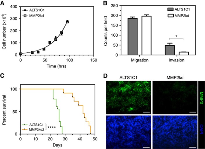
Background: A previous study on a murine astrocytoma cell-line ALTS1C1 showed a highly invasive pattern similar to clinical anaplastic astrocytoma in vivo. This cell-line also expressed a high level of matrix metalloproteinase 2 (MMP2). This study aimed to verify the role of MMP2 in brain tumour progression.
Methods: ALTS1C1 and MMP2 knockdown (MMP2kd) cells were inoculated intracranially, and tumour microenvironment was assessed by immunohistochemistry staining.
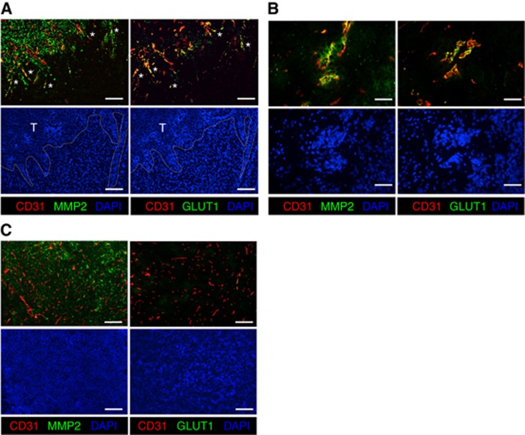
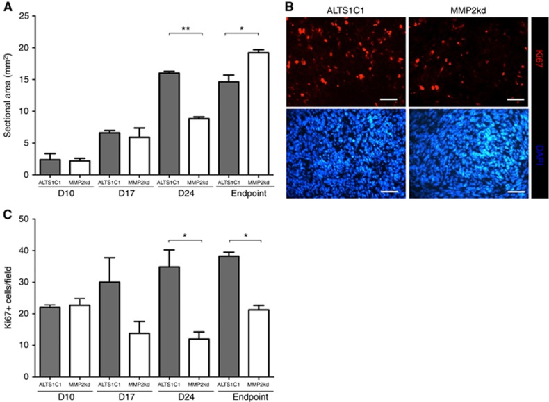
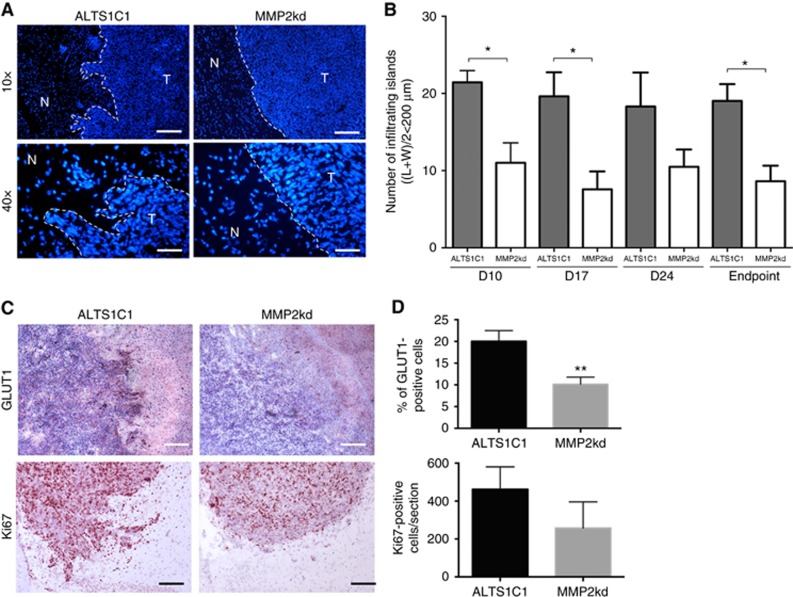
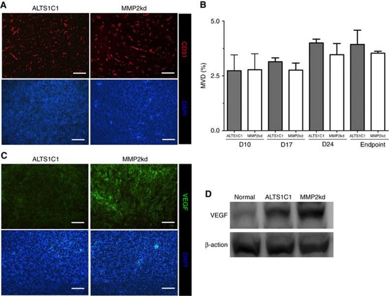
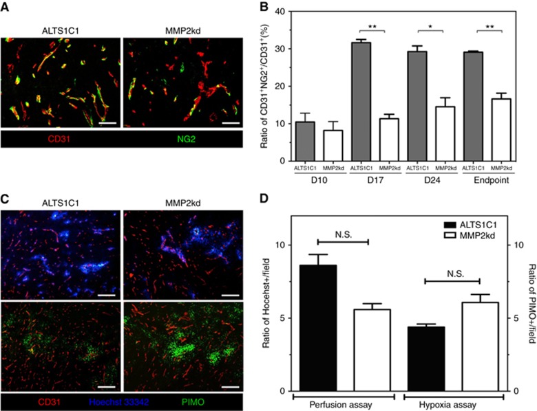
Results: MMP2 expression was co-localised with CD31-positive cells at invading the tumour front and correlated with an invasive marker GLUT-1. The suppression of MMP2 expression prolonged the survival of tumour-bearing mice associated with tumours having smoother tumour margins, decreased Ki67-proliferating index, and down-regulated GLUT-1 antigen. Although the reduction of MMP2 expression did not alter the vessel density in comparison to parental ALTS1C1 tumours, vessels in MMP2kd tumours were less functional, as evidenced by the low ratio of pericyte coverage and reduction in Hoechst33342 dye perfusion.
Conclusions: This study illustrated that tumour-derived MMP2 has at least two roles in tumour malignancy; to enhance tumour invasiveness by degrading the extracellular matrix and to enhance tumour growth by promoting vessel maturation and function.
Conflict of interest statement
The authors declare no conflict of interest.
Figures
Similar articles
-
Matrix metalloproteinase 2 attenuates brain tumour growth, while promoting macrophage recruitment and vascular repair.J Pathol. 2011 Jun;224(2):222-33. doi: 10.1002/path.2854. Epub 2011 Apr 1. J Pathol. 2011. PMID: 21462187
-
PAX6 suppresses the invasiveness of glioblastoma cells and the expression of the matrix metalloproteinase-2 gene.Cancer Res. 2006 Oct 15;66(20):9809-17. doi: 10.1158/0008-5472.CAN-05-3877. Cancer Res. 2006. PMID: 17047041
-
Stromelysin-1/matrix metalloproteinase-3 (MMP-3) expression accounts for invasive properties of human astrocytoma cell lines.Int J Cancer. 2003 Sep 20;106(5):676-82. doi: 10.1002/ijc.11286. Int J Cancer. 2003. PMID: 12866026
-
Effects of pre-irradiation and SDF-1 suppression on the progression of murine astrocytoma cells grown in different stromal beds.Int J Radiat Biol. 2014 Dec;90(12):1162-8. doi: 10.3109/09553002.2014.930539. Epub 2014 Jul 7. Int J Radiat Biol. 2014. PMID: 24937369
-
Stromal matrix metalloproteinase 2 regulates collagen expression and promotes the outgrowth of experimental metastases.J Pathol. 2015 Apr;235(5):773-83. doi: 10.1002/path.4493. Epub 2015 Jan 5. J Pathol. 2015. PMID: 25469981 Free PMC article.
Cited by
-
Insulin promotes invasion and migration of KRASG12D mutant HPNE cells by upregulating MMP-2 gelatinolytic activity via ERK- and PI3K-dependent signalling.Cell Prolif. 2019 May;52(3):e12575. doi: 10.1111/cpr.12575. Epub 2019 Mar 5. Cell Prolif. 2019. PMID: 30838710 Free PMC article.
-
The Function of Oncogene B-Cell Lymphoma 6 in the Regulation of the Migration and Invasion of Trophoblastic Cells.Int J Mol Sci. 2020 Nov 9;21(21):8393. doi: 10.3390/ijms21218393. Int J Mol Sci. 2020. PMID: 33182312 Free PMC article.
-
The Activity of Matrix Metalloproteinases (MMP-2, MMP-9) and Their Tissue Inhibitors (TIMP-1, TIMP-3) in the Cerebral Cortex and Hippocampus in Experimental Acanthamoebiasis.Int J Mol Sci. 2018 Dec 19;19(12):4128. doi: 10.3390/ijms19124128. Int J Mol Sci. 2018. PMID: 30572657 Free PMC article.
-
Molecular Alterations Associated with Acquired Drug Resistance during Combined Treatment with Encorafenib and Binimetinib in Melanoma Cell Lines.Cancers (Basel). 2021 Dec 1;13(23):6058. doi: 10.3390/cancers13236058. Cancers (Basel). 2021. PMID: 34885166 Free PMC article.
-
The Complex Role of Matrix Metalloproteinase-2 (MMP-2) in Health and Disease.Int J Mol Sci. 2024 Dec 21;25(24):13691. doi: 10.3390/ijms252413691. Int J Mol Sci. 2024. PMID: 39769454 Free PMC article. Review.
References
-
- Blumenthal DT, Gorlia T, Gilbert MR, Kim MM, Burt Nabors L, Mason WP, Hegi ME, Zhang P, Golfinopoulos V, Perry JR, Hyun Nam D, Erridge SC, Corn BW, Mirimanoff RO, Brown PD, Baumert BG, Mehta MP, van den Bent MJ, Reardon DA, Weller M, Stupp R (2017) Is more better? The impact of extended adjuvant temozolomide in newly diagnosed glioblastoma: a secondary analysis of EORTC and NRG Oncology/RTOG. Neuro Oncol 19(8): 1119–1126. - PMC - PubMed
-
- Bredin CG, Liu Z, Klominek J (2003) Growth factor-enhanced expression and activity of matrix metalloproteases in human non-small cell lung cancer cell lines. Anticancer Res 23(6C): 4877–4884. - PubMed
-
- Burrell K, Singh S, Jalali S, Hill RP, Zadeh G (2014) VEGF regulates region-specific localization of perivascular bone marrow-derived cells in glioblastoma. Cancer Res 74(14): 3727–3739. - PubMed
-
- Coussens LM, Werb Z (1996) Matrix metalloproteinases and the development of cancer. Chem Biol 3(11): 895–904. - PubMed
MeSH terms
Substances
LinkOut - more resources
Full Text Sources
Other Literature Sources
Medical
Miscellaneous
