. 2018 Jan;118(1):79-87.
doi: 10.1038/bjc.2017.366. Epub 2017 Oct 24.

Identification of a specific peptide binding to colon cancer cells from a phage-displayed peptide library

Affiliations

Identification of a specific peptide binding to colon cancer cells from a phage-displayed peptide library

Lidan Hou et al. Br J Cancer. 2018 Jan.

Abstract

Background: New molecular probes are essential for early colon cancer diagnosis. A phage-display screening was performed to select novel binding peptides for early colon cancer imaging detection.

Methods: A human colon cancer cell line (COLO320HSR) and a normal human intestinal epithelial cell line (NCM460) were used for subtractive screening with a phage peptide library. The positive peptides were identified, and their binding capacities were confirmed by confocal immunofluorescence both in human colon cancer cells and in biopsy specimens. The sequences were further analysed for homology and the existing mimotopes by the BLAST algorithm and the MimoDB database.

Results: A peptide termed as CBP-DWS, which was demonstrated to be capable of binding to a panel of human colon cancer cell lines and tissues, was identified; it had virtually no binding to normal human intestinal epithelial cell line NCM460 and normal surrounding colon tissues. Bioinformatics analyses suggest that CBP-DWS targets human Glypican-3, which may be involved in important cellular functions in multiple cancer types.

Conclusions: These studies suggest that the selected peptide CBP-DWS may be a candidate to serve as a novel probe for colon cancer imaging.


Conflict of interest statement

The authors declare no conflict of interest.

Figures

Figure 1
Figure 1
Selection of specific peptides to COLO320HSR cells by phage display. (A) Schematic representation of M13 filamentous phage. Random foreign DNA fragments are inserted into the phage genomes. The random peptide is shown fused to the amino terminus of the pIII coat protein by a short spacer (Gly-Gly-Gly-Ser, GGGS). (B) Life cycle of non-lytic filamentous phages in host bacteria. The binding of pIII to the bacteria cell membrane leads to injection of phage ssDNA into the bacteria. New phage DNA and proteins are synthesised and then phages are assembled. (C) Workflow of phage-display-based screening in vitro. After four rounds of screening on human colon cancer cells COLO320HSR plus a subtractive screening on normal human intestinal epithelial cells NCM460, cancer cell-bound phages were isolated and sequenced. A full colour version of this figure is available at the British Journal of Cancer journal online.
Figure 2
Figure 2
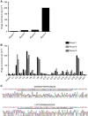
Specific binding of the positive phage clones to COLO320HSR cells. (A) The titres of recovered phages from each round were evaluated by blue plaque-forming assay on an agar plate containing tetracycline. The phage enrichment rate was calculated as output number/input number. (B) After four rounds of biopanning, 21 plaques were randomly selected and analysed by repeated screening on COLO320HSR cells. Four phage clones (C2, C5, C8, C19) appeared to have higher specific binding ability than other clones and were considered as positive phage clones. The unselected original phages were used as negative controls. (C) Amino acid sequence analysis of phage positive clones. The DNA sequences of positive phage clones were sequenced and translated into peptide sequences according to the triple-code theory. A full colour version of this figure is available at the British Journal of Cancer journal online.
Figure 3
Figure 3
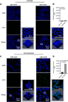
CBP- DWS shows selective homing to COLO320HSR cells. Fixed human colon cancer COLO320HSR cells and normal human intestinal epithelial NCM460 cells were stained with 10 μM l−1 FITC-CBP for 30 min. Strong fluorescent signals were observed on COLO320HSR cells while quite few was detected on NCM460 cells (A, C). Images are also presented in a 2.5-dimension format. The IOD was analysed by Image-Pro Plus 6.0 (B, D). Green colour is from FITC for CBP-DWS. Blue colour is from DAPI for visualising cell nuclei. Scale bar 50 μm. A full colour version of this figure is available at the British Journal of Cancer journal online.
Figure 4
Figure 4
Fluorescent imaging of CBP-DWS binding to a panel of human colon cancer cells. (A) The FITC-labelled peptide CBP-DWS bound to a panel of human colon cancer cells HCT116, HT29, SW480, LoVo, whereas the FITC-labelled control peptide CBP-SCR bound with low affinity to them. Images are also presented in a 2.5-dimension format. Scale bar 20 μm. (B) The IOD of CBP-DWS was significantly higher than that of CBP-SCR. The binding efficiency of the fluorescent peptide to HCT116 (C) and LoVo cells (D) were determined. (E) Unlabelled peptide CBP-DWS were used for competitive inhibition of FITC-CBP-DWS. The fluorescence intensity of FITC-CBP-DWS decreased on both HCT116 and LoVo cells. Scale bar 50 μm. A full colour version of this figure is available at the British Journal of Cancer journal online.
Figure 5
Figure 5
Ex vivo peptide binding to human colon specimens. (A) Frozen serial sections of matched human colon cancer and adjacent normal tissues were stained with 20 μM FITC-CBP-SCR or FITC-CBP-DWS at room temperature for 30 min (n=6). Green colour is from FITC, and blue colour from DAPI for nuclear visualisation. Tissue sections were also stained with H&E and imaged with a digital light microscope. Magnification, × 200. Scale bars: 50 μm. (B) Fluorescence signal intensity analysis reveals CBP-DWS had a significantly higher affinity for colon cancer tissues than CBP-SCR (P=0.0094). A full colour version of this figure is available at the British Journal of Cancer journal online.
Figure 6
Figure 6
Immunofluorescence staining of glypican-3 without and with CBP-DWS blocking. Glypican-3 was detected in HCT116 and LoVo cell lines using sheep anti-human Glypican-3 at 4 °C overnight. And then cells were stained using NorthernLightsTM 557 conjugated anti-sheep secondary antibody (red) and counterstained with DAPI (blue). Bar, 50 μm. A full colour version of this figure is available at the British Journal of Cancer journal online.

References

    1. Bazan J, Calkosinski I, Gamian A (2012) Phage display—a powerful technique for immunotherapy: 1. Introduction and potential of therapeutic applications. Hum Vacc Immunother 8(12): 1817–1828. - PMC - PubMed
    1. Choi Y, Sateia HF, Peairs KS, Stewart RW (2017) Screening for colorectal cancer. Semin Oncol 44(1): 34–44. - PubMed
    1. De Robertis M, Arigoni M, Loiacono L, Riccardo F, Calogero RA, Feodorova Y, Tashkova D, Belovejdov V, Sarafian V, Cavallo F, Signori E (2015) Novel insights into Notum and glypicans regulation in colorectal cancer. Oncotarget 6(38): 41237–41257. - PMC - PubMed
    1. Filmus J, Capurro M (2013) Glypican-3: a marker and a therapeutic target in hepatocellular carcinoma. FEBS J 280(10): 2471–2476. - PubMed
    1. Filmus J, Capurro M, Rast J (2008) Glypicans. Genome Biol 9(5): 224. - PMC - PubMed

Publication types