

Zinc-finger protein 418 overexpression protects against cardiac hypertrophy and fibrosis
- PMID: 29065170
- PMCID: PMC5655480
- DOI: 10.1371/journal.pone.0186635
Zinc-finger protein 418 overexpression protects against cardiac hypertrophy and fibrosis
Abstract
Background: This study aimed to investigated the effect and mechanism of zinc-finger protein 418 (ZNF418) on cardiac hypertrophy caused by aortic banding (AB), phenylephrine (PE) or angiotensin II (Ang II) in vivo and in vitro.
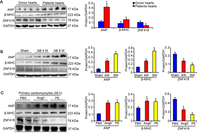
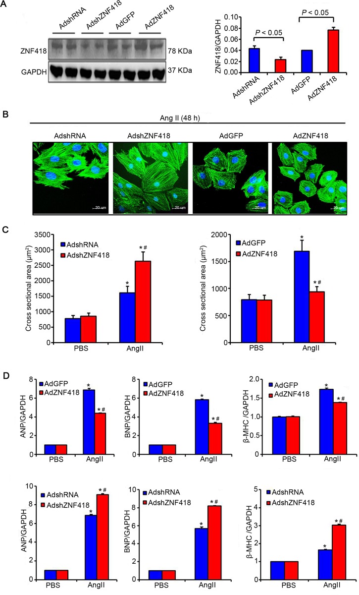
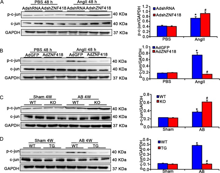
Methods: The expression of ZNF418 in hearts of patients with dilated cardiomyopathy (DCM) or hypertrophic cardiomyopathy (HCM) and AB-induced cardiac hypertrophy mice, as well as in Ang II- or PE-induced hypertrophic primary cardiomyocytes was detected by western blotting. Then, the expression of ZNF418 was up-regulated or down-regulated in AB-induced cardiac hypertrophy mice and Ang II -induced hypertrophic primary cardiomyocytes. The hypertrophic responses and fibrosis were evaluated by echocardiography and histological analysis. The mRNA levels of hypertrophy markers and fibrotic markers were detected by RT-qPCR. Furthermore, the phosphorylation and total levels of c-Jun were measured by western blotting.
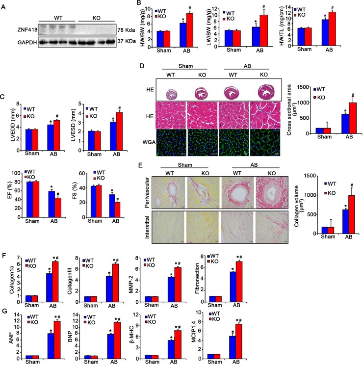
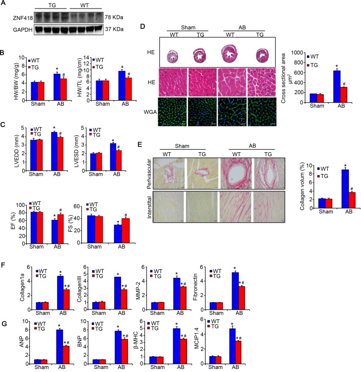
Results: ZNF418 was markedly down-regulated in hearts of cardiac hypertrophy and hypertrophic primary cardiomyocytes. Down-regulated ZNF418 exacerbated the myocyte size and fibrosis, moreover increased the mRNA levels of ANP, BNP, β-MHC, MCIP1.4, collagen 1a, collagen III, MMP-2 and fibronection in hearts of AB-treated ZNF418 knockout mice or Ang II-treated cardiomyocytes with AdshZNF418. Conversely, these hypertrophic responses were reduced in the ZNF418 transgenic (TG) mice treated by AB and the AdZNF418-transfected primary cardiomyocytes treated by Ang II. Additionally, the deficiency of ZNF418 enhanced the phosphorylation level of c-jun, and overexpression of ZNF418 suppressed the phosphorylation level of c-jun in vivo and in vitro.
Conclusion: ZNF418 maybe attenuate hypertrophic responses by inhibiting the activity of c-jun/AP-1.
Conflict of interest statement
Figures
Similar articles
-
GDF11 Attenuated ANG II-Induced Hypertrophic Cardiomyopathy and Expression of ANP, BNP and Beta-MHC Through Down- Regulating CCL11 in Mice.Curr Mol Med. 2018;18(10):661-671. doi: 10.2174/1566524019666190204112753. Curr Mol Med. 2018. PMID: 30714521
-
Effect of endostatin overexpression on angiotensin II-induced cardiac hypertrophy in rats.Chin Med J (Engl). 2019 Nov 20;132(22):2716-2723. doi: 10.1097/CM9.0000000000000513. Chin Med J (Engl). 2019. PMID: 31725448 Free PMC article.
-
Downregulation of salusins alleviates hypertrophic cardiomyopathy via attenuating oxidative stress and autophagy.Eur J Med Res. 2024 Feb 9;29(1):109. doi: 10.1186/s40001-024-01676-z. Eur J Med Res. 2024. PMID: 38336819 Free PMC article.
-
JunD attenuates phenylephrine-mediated cardiomyocyte hypertrophy by negatively regulating AP-1 transcriptional activity.Cardiovasc Res. 2006 Jul 1;71(1):108-17. doi: 10.1016/j.cardiores.2006.02.032. Epub 2006 Mar 7. Cardiovasc Res. 2006. PMID: 16690042
-
A novel inhibitory role for CREG-mediated signalling in cardiac hypertrophy?J Hypertens. 2004 Aug;22(8):1469-71. doi: 10.1097/01.hjh.0000133741.00738.a2. J Hypertens. 2004. PMID: 15257166 Review. No abstract available.
Cited by
-
ZNF667 facilitates angiogenesis after myocardial ischemia through transcriptional regulation of VASH1 and Wnt signaling pathway.Int J Mol Med. 2022 Oct;50(4):129. doi: 10.3892/ijmm.2022.5185. Epub 2022 Aug 31. Int J Mol Med. 2022. PMID: 36043524 Free PMC article.
-
miR-1204 promotes hepatocellular carcinoma progression through activating MAPK and c-Jun/AP1 signaling by targeting ZNF418.Int J Biol Sci. 2019 Jun 2;15(7):1514-1522. doi: 10.7150/ijbs.33658. eCollection 2019. Int J Biol Sci. 2019. Retraction in: Int J Biol Sci. 2022 Jun 7;18(10):3942. doi: 10.7150/ijbs.75583. PMID: 31337980 Free PMC article. Retracted.
-
Identification of biomarkers, pathways, and potential therapeutic targets for heart failure using next-generation sequencing data and bioinformatics analysis.Ther Adv Cardiovasc Dis. 2023 Jan-Dec;17:17539447231168471. doi: 10.1177/17539447231168471. Ther Adv Cardiovasc Dis. 2023. PMID: 37092838 Free PMC article.
-
Pharmacological Inhibition of Caspase-1 Ameliorates Cisplatin-Induced Nephrotoxicity through Suppression of Apoptosis, Oxidative Stress, and Inflammation in Mice.Mediators Inflamm. 2018 Dec 23;2018:6571676. doi: 10.1155/2018/6571676. eCollection 2018. Mediators Inflamm. 2018. PMID: 30670928 Free PMC article.
-
Cooperative regulation of Zhx1 and hnRNPA1 drives the cardiac progenitor-specific transcriptional activation during cardiomyocyte differentiation.Cell Death Discov. 2023 Jul 14;9(1):244. doi: 10.1038/s41420-023-01548-1. Cell Death Discov. 2023. PMID: 37452012 Free PMC article.
References
-
- Shimizu I, Minamino T (2016) Physiological and pathological cardiac hypertrophy. Journal of molecular and cellular cardiology. - PubMed
-
- Maron BJ, Maron MS (2013) Hypertrophic cardiomyopathy. The Lancet 381: 242–255. - PubMed
-
- Finckenberg P, Mervaala E (2010) Novel regulators and drug targets of cardiac hypertrophy. Journal of hypertension 28: S33–S38. doi: 10.1097/01.hjh.0000388492.73954.0b - DOI - PubMed
-
- Eder P, Molkentin JD (2011) TRPC channels as effectors of cardiac hypertrophy. Circulation research 108: 265–272. doi: 10.1161/CIRCRESAHA.110.225888 - DOI - PubMed
-
- Ling H, Zhang T, Pereira L, Means CK, Cheng H, Gu Y, et al. (2009) Requirement for Ca 2+/calmodulin–dependent kinase II in the transition from pressure overload–induced cardiac hypertrophy to heart failure in mice. The Journal of clinical investigation 119: 1230–1240. doi: 10.1172/JCI38022 - DOI - PMC - PubMed
MeSH terms
Substances
LinkOut - more resources
Full Text Sources
Other Literature Sources
Molecular Biology Databases
Research Materials
Miscellaneous
