

Comparative analysis of activation induced marker (AIM) assays for sensitive identification of antigen-specific CD4 T cells
- PMID: 29065175
- PMCID: PMC5655442
- DOI: 10.1371/journal.pone.0186998
Comparative analysis of activation induced marker (AIM) assays for sensitive identification of antigen-specific CD4 T cells
Abstract
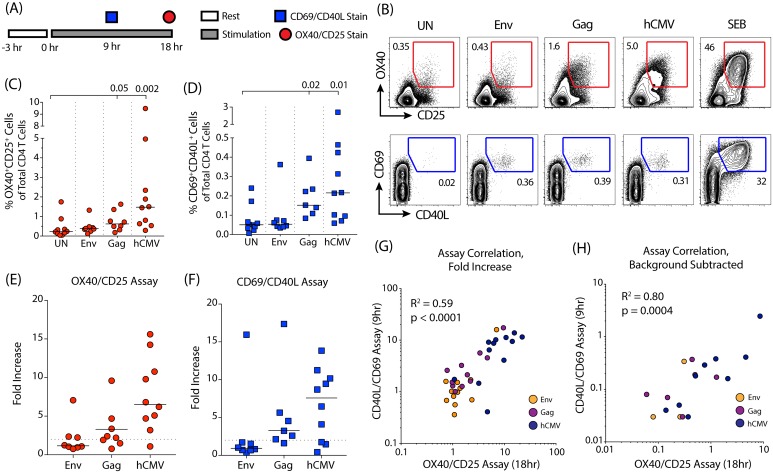
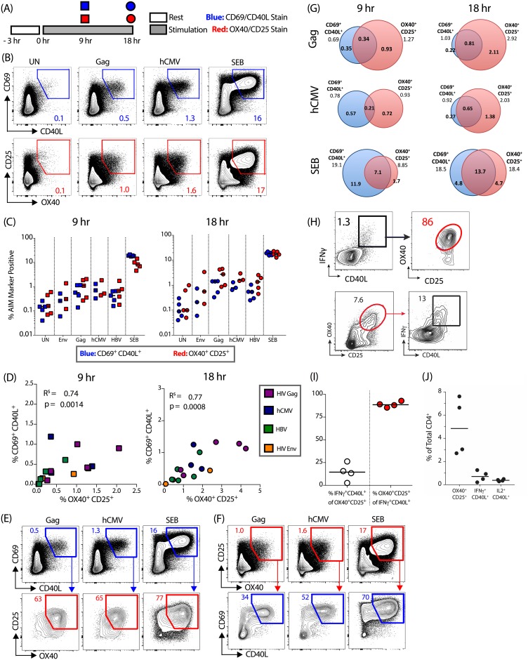
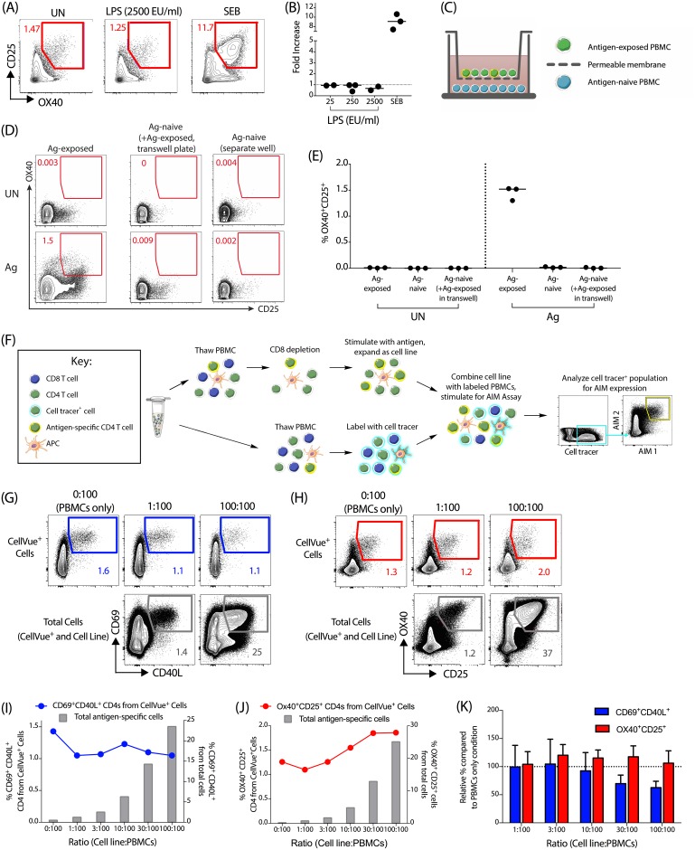
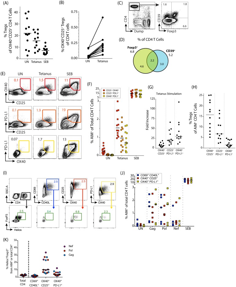
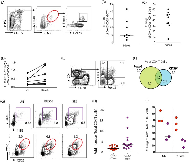
The identification and study of antigen-specific CD4 T cells, both in peripheral blood and in tissues, is key for a broad range of immunological research, including vaccine responses and infectious diseases. Detection of these cells is hampered by both their rarity and their heterogeneity, in particular with regards to cytokine secretion profiles. These factors prevent the identification of the total pool of antigen-specific CD4 T cells by classical methods. We have developed assays for the highly sensitive detection of such cells by measuring the upregulation of surface activation induced markers (AIM). Here, we compare two such assays based on concurrent expression of CD69 plus CD40L (CD154) or expression of OX40 plus CD25, and we develop additional AIM assays based on OX40 plus PD-L1 or 4-1BB. We compare the relative sensitivity of these assays for detection of vaccine and natural infection-induced CD4 T cell responses and show that these assays identify distinct, but overlapping populations of antigen-specific CD4 T cells, a subpopulation of which can also be detected on the basis of cytokine synthesis. Bystander activation had minimal effect on AIM markers. However, some T regulatory cells upregulate CD25 upon antigen stimulation. We therefore validated AIM assays designed to exclude most T regulatory cells, for both human and non-human primate (NHP, Macaca mulatta) studies. Overall, through head-to-head comparisons and methodological improvements, we show that AIM assays represent a sensitive and valuable method for the detection of antigen-specific CD4 T cells.
Conflict of interest statement
Figures
References
-
- Horton H, Thomas EP, Stucky JA, Frank I, Moodie Z, Huang Y, et al. Optimization and validation of an 8-color intracellular cytokine staining (ICS) assay to quantify antigen-specific T cells induced by vaccination. J Immunol Methods. 2007;323: 39–54. doi: 10.1016/j.jim.2007.03.002 - DOI - PMC - PubMed
-
- Lamoreaux L, Roederer M, Koup R. Intracellular cytokine optimization and standard operating procedure. Nat Protoc. 2006;1: 1507–1516. doi: 10.1038/nprot.2006.268 - DOI - PubMed
-
- Sallusto F, Cassotta A, Hoces D, Foglierini M, Lanzavecchia A. Do Memory CD4 T Cells Keep Their Cell-Type Programming: Plasticity versus Fate Commitment? Cold Spring Harb Perspect Biol. 2017;: a029421–10. doi: 10.1101/cshperspect.a029421 - DOI - PMC - PubMed
-
- Crotty S. Do Memory CD4 T Cells Keep Their Cell-Type Programming: Plasticity versus Fate Commitment? Cold Spring Harb Perspect Biol. 2017;: a032102–13. doi: 10.1101/cshperspect.a032102 - DOI - PMC - PubMed
-
- Kroenke MA, Eto D, Locci M, Cho M, Davidson T, Haddad EK, et al. Bcl6 and Maf cooperate to instruct human follicular helper CD4 T cell differentiation. The Journal of Immunology. 2012;188: 3734–3744. doi: 10.4049/jimmunol.1103246 - DOI - PMC - PubMed
Publication types
MeSH terms
Substances
Grants and funding
LinkOut - more resources
Full Text Sources
Other Literature Sources
Research Materials
