. 2018 Jan;175(2):374-387.
doi: 10.1111/bph.14079. Epub 2017 Dec 15.

Berberine attenuates hepatic steatosis and enhances energy expenditure in mice by inducing autophagy and fibroblast growth factor 21

Affiliations

Berberine attenuates hepatic steatosis and enhances energy expenditure in mice by inducing autophagy and fibroblast growth factor 21

Yixuan Sun et al. Br J Pharmacol. 2018 Jan.

Abstract

Background and purpose: Berberine, a compound from rhizome coptidis, is traditionally used to treat gastrointestinal infections, such as bacterial diarrhoea. Recently, berberine was shown to have hypoglycaemic and hypolipidaemic effects. We investigated the mechanisms by which berberine regulates hepatic lipid metabolism and energy expenditure in mice.

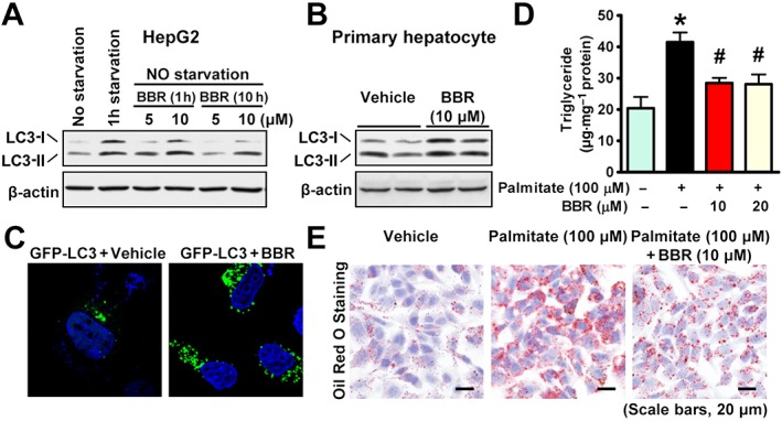
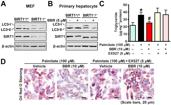
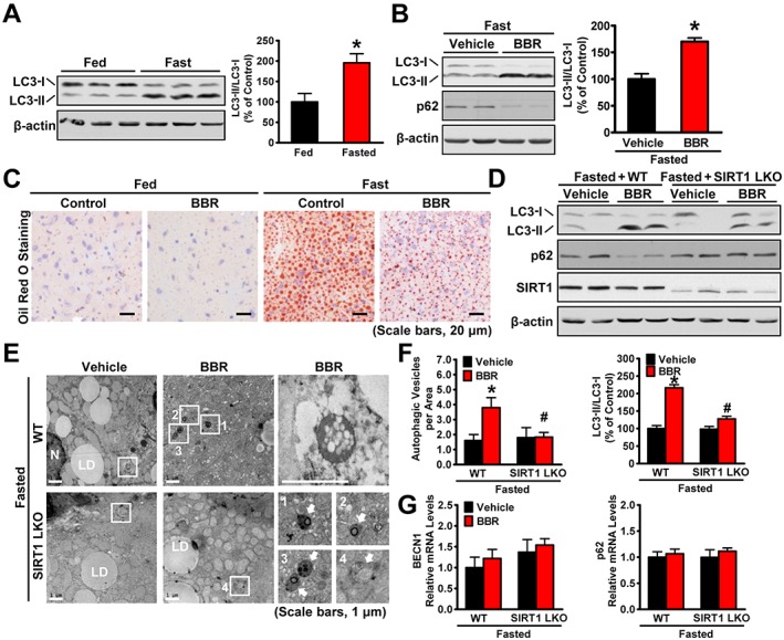
Experimental approach: Liver-specific SIRT1 knockout mice and their wild-type littermates were fed a high-fat, high-sucrose (HFHS) diet and treated with berberine by i.p. injection for five weeks. Mouse primary hepatocytes and human HepG2 cells were treated with berberine and then subjected to immunoblotting analysis and Oil Red O staining.

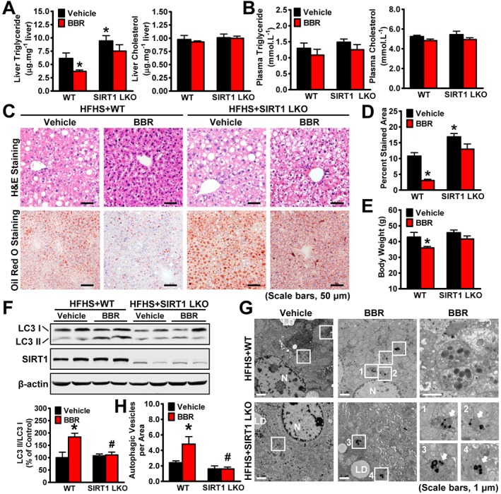
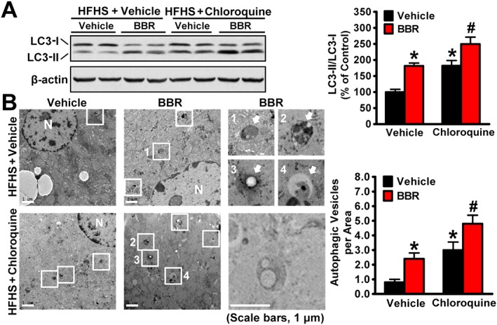
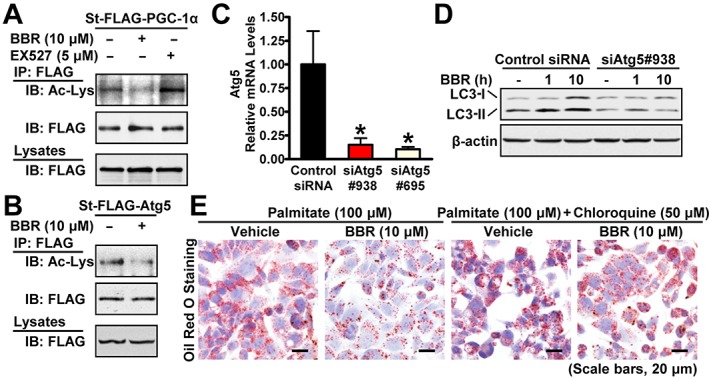
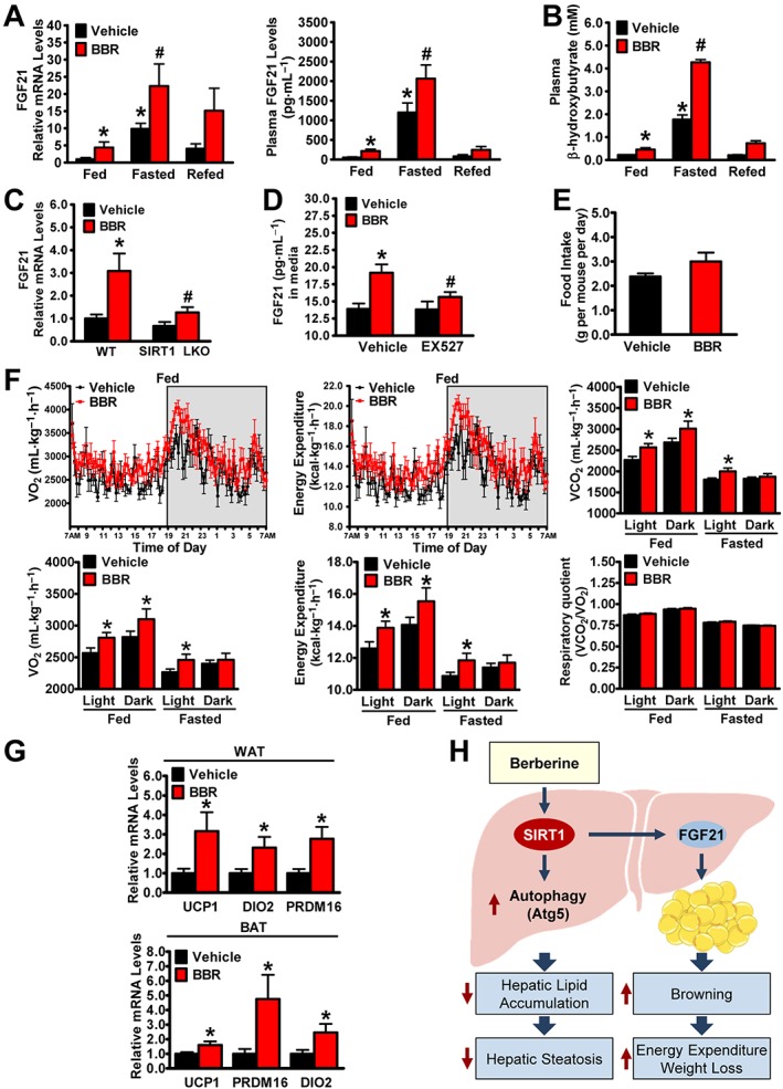
Key results: Berberine attenuated hepatic steatosis and controlled energy balance in mice by inducing autophagy and FGF21. These beneficial effects of berberine on autophagy and hepatic steatosis were abolished by a deficiency of the nutrient sensor SIRT1 in the liver of HFHS diet-fed obese mice and in mouse primary hepatocytes. SIRT1 is essential for berberine to potentiate autophagy and inhibit lipid storage in mouse livers in response to fasting. Mechanistically, the berberine stimulates SIRT1 deacetylation activity and induces autophagy in an autophagy protein 5-dependent manner. Moreover, the administration of berberine was shown to promote hepatic gene expression and circulating levels of FGF21 and ketone bodies in mice in a SIRT1-dependent manner.

Conclusions and implications: Berberine acts in the liver to regulate lipid utilization and maintain whole-body energy metabolism by mediating autophagy and FGF21 activation. Hence, it has therapeutic potential for treating metabolic defects under nutritional overload, such as fatty liver diseases, type 2 diabetes and obesity.


Figures

Figure 1
Figure 1
The beneficial effects of berberine (BBR) on body weight and hepatic steatosis are compromised by hepatic‐specific deletion of SIRT1 in HFHS diet‐fed mice. WT and SIRT1 LKO male mice at 16 weeks old were fed on a HFHS diet for 12 weeks and then treated with berberine (5 mg·kg−1·day−1) or vehicle (PBS) by i.p. injection once daily for 5 weeks. (A) Liver and (B) plasma triglyceride and cholesterol levels were assessed in mice. (C) Representative H&E and Oil Red O staining and (D) quantification of Oil Red O‐stained areas are shown, n = 6. (E) The body weight was measured, n = 6. (F‐H) SIRT1 is necessary for berberine‐induced autophagy in the liver of HFHS diet‐fed mice. (F) Berberine‐stimulated conversion of LC3‐I to LC3‐II is reduced in SIRT1 LKO mice fed a HFHS diet. Band intensity of LC3‐II was quantified by densitometry and normalized to the levels of LC3‐I for unwanted sources of variation and presented as the mean ± SEM, n = 6. *P < 0.05, versus WT and vehicle; #P < 0.05, versus WT mice treated with berberine. (G) Electron micrographs of the liver tissue. Images 1–4 showing AVs are high magnifications of the ‘white box’ area indicated in the images of berberine‐treated WT or SIRT1 LKO mice fed a HFHS diet. Arrows denote autophagic vacuoles (AVs). N, nucleus; LD, lipid droplet. (H) The average number of AVs was quantified from a randomly selected pool of five fields under each condition. The data are presented as the mean ± SEM, n = 5. *P < 0.05, versus WT and vehicle; #P < 0.05, versus WT mice treated with berberine.
Figure 2
Figure 2
Berberine (BBR) is able to induce autophagic flux in the livers of HFHS diet‐fed mice. (A) Eight‐week‐old male C57BL/6 mice were fed on a HFHS diet for 9 weeks and then treated with berberine (5 mg·kg−1·day−1) once daily for 4 weeks, followed by treatment with chloroquine (60 mg·kg−1) i.p. for 24 h before the kill. (A) Berberine stimulates conversion of LC3‐I to LC3‐II under chloroquine treatment in the liver of mice fed with HFHS diet. Band intensity of LC3‐II was quantified by densitometry and normalized to the levels of LC3‐I for unwanted sources of variation and presented as the mean ± SEM, n = 5. *P < 0.05, versus WT and vehicle; #P < 0.05, versus WT mice treated with berberine. (B) Electron micrographs of the liver tissue. Images 1–4 showing AVs are high magnifications of the ‘white box’ area indicated in the images of berberine‐treated or chloroquine‐treated mice fed a HFHS diet. Arrows denote autophagic vacuoles (AVs). N, nucleus. The average numbers of AVs were quantified from a randomly selected pool of five fields under each condition. The data are presented as the mean ± SEM, n = 5. *P < 0.05, versus vehicle; #P < 0.05, versus vehicle and chloroquine.
Figure 3
Figure 3
Berberine (BBR) stimulates autophagy to repress lipid accumulation in hepatocytes. Berberine stimulates the conversion of LC3‐I to LC3‐II in (A) HepG2 cells and (B) primary mouse hepatocytes. (C) The redistribution of GFP‐LC3 to form puncta is enhanced by berberine in HeLa cells. Cells were transfected with expressing plasmid encoding GFP‐LC3 fusion protein, followed by treatment without or with 10 μM berberine for 10 h. Berberine decreased lipid accumulation in HepG2 cells exposed to palmitate, as reflected by triglyceride levels determined by a (D) colorimetric enzymatic assay and (E) Oil Red O staining. The data are presented as the mean ± SEM, n = 6. *P < 0.05, versus control; #P < 0.05, versus palmitate.
Figure 4
Figure 4
Berberine (BBR) induces autophagy to inhibit lipid accumulation through SIRT1 in hepatocytes. (A) Effects of SIRT1 deficiency on LC3 in MEFs. MEFs of floxed SIRT1 mice were infected with adenoviral Cre for 72 h. (B) SIRT1 deficiency diminishes berberine‐stimulated conversion of LC3‐I to LC3‐II in primary hepatocytes. Primary hepatocytes were isolated from 8‐week‐old WT or SIRT1 LKO mice and treated as indicated. The lipid‐lowering effects of berberine were abolished by a pharmacological SIRT1 inhibitor EX527 in HepG2 cells treated with palmitate, as determined by (C) triglyceride levels and (D) Oil Red O staining. The data are presented as the mean ± SEM, n = 6. *P < 0.05, versus control; #P < 0.05, versus palmitate.
Figure 5
Figure 5
Berberine (BBR) stimulates SIRT1 deacetylation activity and induces autophagy in an Atg5‐dependent manner. Berberine induces deacetylation of (A) PGC‐1α or (B) Atg5. HEK293 cells stably expressing mouse FLAG‐tagged PGC‐1α, human FLAG‐tagged Atg5 (St‐FLAG‐Atg5) or empty vector were treated with 10 μM berberine or 5 μM EX527 for 10 h or 6 h, respectively, followed by immunoprecipitation and immunoblots with the indicated antibodies. (C) The levels of mRNA for Atg5 are decreased by Atg5 knockdown in HepG2 cells. The data were normalized to β‐actin for unwanted sources of variation and presented as the mean ± SEM, n = 5. *P < 0.05, versus control siRNA. (D) Berberine‐stimulated conversion of LC3‐I to LC3‐II is abolished by Atg5 knockdown in HepG2 cells. Cells were transfected with siAtg5 and control siRNAs for 24 h and incubated in serum‐free DMEM containing 5.5 mM glucose overnight, followed by treatment without or with 10 μM berberine for 1 h and 10 h. (E) Berberine's lipid‐lowering effects are attenuated by the autophagy inhibitor chloroquine in HepG2 cells treated with palmitate as reflected by Oil Red O staining.
Figure 6
Figure 6
SIRT1 is required for berberine (BBR) to potentiate fasting‐induced autophagy in mouse livers. Male C57BL/6 mice at 8 weeks of age and SIRT1 LKO or WT mice at 16 weeks of age were treated with berberine (5 mg·kg−1·day−1) or vehicle (PBS) by i.p. injection once daily for 4 weeks and then subjected to feeding (fed) or fasting for 24 h (fasted). (A) The conversion of LC3‐I to LC3‐II was increased by fasting in mice. Band intensity of LC3‐II was quantified by densitometry and normalized to the levels of LC3‐I for unwanted sources of variation and presented as the mean ± SEM, n = 5. *P < 0.05, versus fed mice. (B) Berberine stimulates the conversion of LC3‐I to LC3‐II and reduces the p62 in mice in response to fasting, n = 5. (C) Hepatic steatosis was assessed by Oil Red O staining (scale bars: 50 μm). (D) SIRT1 deficiency attenuated berberine‐induced conversion of LC3‐I to LC3‐II and the reduction of p62 in the liver of fasted mice, n = 6. (E) Electron micrographs and the (F) average numbers of autophagic vacuoles (AVs) in the liver tissue were measured. Images 1–4 showing AVs are high magnifications of the ‘white box’ area indicated in the images of berberine‐treated WT or SIRT1 LKO mice in response to fasting. Arrows denote AVs. N, nucleus; LD, lipid droplet. The average numbers of AVs were quantified from a randomly selected pool of five fields under each condition. The data are presented as the mean ± SEM, n = 5. *P < 0.05, versus WT and vehicle; #P < 0.05, versus WT and berberine. (G) The mRNA levels of BECN1 and p62 were measured by real‐time PCR. The data were normalized to β‐actin for unwanted sources of variation and presented as the mean ± SEM, n = 5.
Figure 7
Figure 7
The administration of berberine (BBR) induces the production and secretion of FGF21 and promotes whole‐body energy expenditure in mice. Male C57BL/6 mice at 8 weeks of age were treated with berberine (5 mg·kg−1·day−1) or vehicle (PBS) via i.p. injection for 4 weeks and then subjected to feeding (fed), fasting for 24 h (fasted) or refeeding for 6 h after a 24 h fast (refed). (A) Administration of berberine is able to promote hepatic gene expression and circulating levels of FGF21 in mice. The mRNA levels were normalized to β‐actin for unwanted sources of variation, n = 5. (B) Circulating ketone bodies levels were robustly induced by treatment with berberine. The data are presented as the mean ± SEM, n = 5. *P < 0.05, versus fed and vehicle; #P < 0.05, versus fasted and vehicle. (C) SIRT1 deficiency abolishes berberine‐induced expression of FGF21 in mouse primary hepatocytes. The mRNA levels were normalized to β‐actin for unwanted sources of variation and presented as the mean ± SEM, n = 6. *P < 0.05, versus WT and vehicle; #P < 0.05, versus WT and berberine. (D) Effects of SIRT1 inhibitor EX527 on FGF21 secretion in hepatocytes. Media FGF21 levels were measured in HepG2 cells treated with berberine (10 μM) and EX527 (5 μM). The data are presented as the mean ± SEM, n = 5. *P < 0.05, versus vehicle; #P < 0.05, versus vehicle and berberine. (E) Food intake was measured during the fed state. (F) The rate of VO2, energy expenditure, VCO2 and respiratory quotient were measured by comprehensive metabolic monitoring in mice over a 24 h period with food and over a 24 h fast and normalized to lean body mass. (G) Administration of berberine increases the transcription of brown‐like genes in WAT and BAT in mice. The mRNA levels were normalized to β‐actin for unwanted sources of variation and presented as the mean ± SEM, n = 8. *P < 0.05, versus vehicle. (H) The proposed model for SIRT1 in mediating berberine's amelioration of hepatic steatosis and obesity in diet‐induced insulin‐resistant mice. Administration of berberine stimulates hepatic SIRT1, which in turn increases autophagic function to regulate intracellular lipid stores, and improves fatty liver disease. Activation of the berberine‐SIRT1‐FGF21 axis may represent a new approach to treat obesity by promoting white fat browning and stimulating energy expenditure.

References

    1. Ahmadian M, Suh JM, Hah N, Liddle C, Atkins AR, Downes M et al (2013). PPARγ signaling and metabolism: the good, the bad and the future. Nat Med 99: 557–566. - PMC - PubMed
    1. Alexander SPH, Fabbro D, Kelly E, Marrion NV, Peters JA, Faccenda E et al (2017a). The Concise Guide to PHARMACOLOGY 2017/18: Enzymes. Br J Pharmacol 174: S272–S359. - PMC - PubMed
    1. Alexander SPH, Kelly E, Marrion NV, Peters JA, Faccenda E, Harding SD et al (2017b). The Concise Guide to PHARMACOLOGY 2017/18: Transporters. Br J Pharmacol 174: S360–S446. - PMC - PubMed
    1. Badman MK, Pissios P, Kennedy AR, Koukos G, Flier JS, Maratos‐Flier E (2007). Hepatic fibroblast growth factor 21 is regulated by PPARα and is a key mediator of hepatic lipid metabolism in ketotic states. Cell Metab 5: 426–437. - PubMed
    1. Baur JA, Pearson KJ, Price NL, Jamieson HA, Lerin C, Kalra A et al (2006). Resveratrol improves health and survival of mice on a high‐calorie diet. Nature 444: 337–342. - PMC - PubMed

Publication types

MeSH terms