Review
. 2017 Dec;6(24):10.1002/adhm.201700681.
doi: 10.1002/adhm.201700681. Epub 2017 Oct 24.

Fundamentals of Laser-Based Hydrogel Degradation and Applications in Cell and Tissue Engineering

Affiliations
Review

Fundamentals of Laser-Based Hydrogel Degradation and Applications in Cell and Tissue Engineering

Shantanu Pradhan et al. Adv Healthc Mater. 2017 Dec.

Abstract

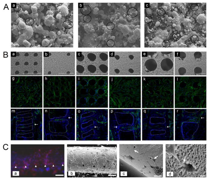
The cell and tissue engineering fields have profited immensely through the implementation of highly structured biomaterials. The development and implementation of advanced biofabrication techniques have established new avenues for generating biomimetic scaffolds for a multitude of cell and tissue engineering applications. Among these, laser-based degradation of biomaterials is implemented to achieve user-directed features and functionalities within biomimetic scaffolds. This review offers an overview of the physical mechanisms that govern laser-material interactions and specifically, laser-hydrogel interactions. The influences of both laser and material properties on efficient, high-resolution hydrogel degradation are discussed and the current application space in cell and tissue engineering is reviewed. This review aims to acquaint readers with the capability and uses of laser-based degradation of biomaterials, so that it may be easily and widely adopted.

Keywords: biofabrication; biomaterials; biomimetics; cell migration; microfluidics; microphysiological systems; neuronal guidance.


Figures

Figure 1
Figure 1. Overview of laser-based degradation in literature
(A) There has been a steady increase in the number of publications involving laser-based degradation of hydrogels and biomaterials over the past two decades. (B) Trends in the use of different biomaterials manipulated via laser-based degradation. (C) Categorization of publications based on biomaterial type and (D) application. The graphs represent (A, B) the number of publications or (C, D) percentages, that contain the terms ‘laser degradation’, ‘laser structuring’, ‘laser micropatterning’, ‘laser ablation’, ‘hydrogels’, and ‘scaffolds’ from searches using NCBI Pubmed, Google Scholar, and the ISI Web of Knowledge.
Figure 2
Figure 2. Theoretical power and intensity outputs for three different lasers over time as a function of pulse duration and pulse frequency
(A) Comparison of the power and intensity over time for three different lasers (continuous wave, 1 ns pulsed, and 140 fs pulsed) operating at the same pulse frequency for the pulsed lasers (80 MHz), same average power (1 W), and same average intensity (1.34×10⁸ W.cm−2) but with different pulse durations, 1 ns and 140 fs for the pulsed lasers. The fs pulsed laser outputs a higher peak power and intensity compared to the ns pulsed or continuous wave lasers to achieve the same average power and intensity. (B) Comparison of the power and intensity output over time for three different lasers (continuous wave, low frequency (8 MHz) fs pulsed, and high frequency (80 MHz) fs pulsed) operating at the same average power (1 W) and same average intensity (1.34×10⁸ W.cm−2) with the same pulse duration for the pulsed lasers (140 fs) but at different frequencies. The laser operating at the lower frequency (8 MHz) outputs a higher peak power and intensity to achieve the same average output compared to the higher frequency pulse and continuous wave lasers. (IP: peak intensity, PP: peak power, IAVG: average intensity, PAVG: average power).
Figure 3
Figure 3. Interplay of physical mechanisms influencing laser-based degradation
(A) The sequential occurrence of photoionization, inverse Bremsstrahlung absorption, and impact ionization leads to plasma formation and recurring sequences in absorption and ionization events lead to avalanche growth of free electrons produced by laser excitation. Reproduced with permission.[48] Copyright 2005, Springer-Verlag. (B) Schematic illustration of (a) distribution of processes in the focal volume and (b) sequence of processes occurring over the course of a laser pulse. Zener ionization and Zener-seeded avalanche ionization cause increases in the electron density and plasma field intensity in the focal volume. The laser penetration depth (δ) becomes approximately equal to the wavelength (λ) at 1021 electrons/cm3. When plasma frequency (ωp) becomes equal to the laser frequency (ω), rapid ionization and heavy absorption take place and material over depth, δ, is vaporized. The pink region at the bottom indicates the duration of the laser pulse. EQ and EG refer to the electron quiver energy and the band gap respectively. Reproduced with permission.[55] Copyright 2004, The National Academy of Sciences of the USA. (C) (a) Overview of the range of laser-tissue interactions that occur at varying energy density, power density, and pulse duration and (b) inverse variation of threshold fluence with absorption coefficient over a range of wavelengths. (b) Solid black line indicates the absorption coefficient of water (cm−1), dashed line connecting solid filled circles indicates the absorption coefficient of tissue (cm−1), and dashed line connecting hollow circles indicates the threshold fluence for laser ablation (mJ.mm−2). Reproduced with permission.[50] Copyright 1991, Baillière Tindall.
Figure 4
Figure 4. Effect of pulse duration on laser-material interactions
(A) (a) Degradation using a femtosecond pulsed laser provides increased spatial resolution compared to a nanosecond pulsed as demonstrated by the lateral and axial dimensions of the degradation volume. (b) The visible laser-induced damage proportional to the light intensity (ΔI) as a function of the peak laser intensity for nanosecond and femtosecond pulsed lasers. (c) The measured (X symbols) and theoretical (curves) ablation threshold values versus pulse duration for degradation of PEG-fibrinogen hydrogels. (B) Comparison of visible damage within PEG-fibrinogen hydrogels caused by nanosecond and femtosecond pulsed lasers as a function of laser intensity. Scale bar = 100 μm. (A–B) Reproduced with permission.[31] Copyright 2009, The Biophysical Society. (C) Plasma, shock wave, and cavitation bubble formation in water produced by Nd:YAG laser pulses of different pulse duration and energy, imaged 44 ns after the optical breakdown. Scale bar = 100 μm. Reproduced with permission.[58] Copyright 1996, Acoustical Society of America.
Figure 5
Figure 5. Characterization of the two-photon excitation volume
(A) Visualization of the excitation volume for single-photon (1P) and two-photon (2P) excitation of fluorescein using (a) a continuous wave laser at 488 nm and (b) a femtosecond pulsed laser at 960 nm focused through a NA 0.16 objective. (B) (a) Lateral and axial views of the point spread function using 1P and 2P excitation. (b) FWHM refers to the full-width half-maximum of the Gaussian fit and ω refers to the axial radius. (C) The 2P excitation volume calculated for a 1-GM and a 300-GM fluorophore excited using a 200 fs pulsed laser operating at 80 MHz focused through a 1.2 NA objective. Inset shows the point spread function for excitation of a 1 GM (left) and 300 GM (right) fluorophore at 20 mW. (A–C) Reproduced with permission.[59] Copyright 2003, Nature Publishing Group. (D) The distribution of the normalized intensity and electron density in a focal volume of a femtosecond pulsed laser during optical breakdown using a 1.3 NA objective and 800 nm light. Reproduced with permission.[48] Copyright 2005, Springer-Verlag.
Figure 6
Figure 6. Schematic of the various modes of laser-hydrogel interactions
Laser-induced degradation of hydrogels can follow various modes depending on the type and composition of polymeric material. Scheme 1: the water within the polymeric network is excited by the laser and undergoes electron generation, plasma formation, and vapor bubble generation. The vapor bubble expands radially along with shock wave propagation and heat dissipation and subsequently causes physical breakage of the network due to thermoelastic stress. Scheme 2: the polymeric network is excited by the incident laser light and undergoes plasma formation which subsequently leads to physical breakage of polymer network. Scheme 3: hydrogels with a high two-photon cross-section or those containing photolabile groups undergo direct chemical scission which induces rapid dissociation of the polymer network.
Figure 7
Figure 7. Neuronal guidance using laser-degraded networks
(A) Degradation of microchannels between agar microchambers enables the formation of functional connections between rat hippocampal cells. (a) Microchannels (indicated by white arrows) are fabricated 1 day after cells are introduced, (b) cellular connections are formed over 5 days, (c) photothermal etching to create new channels in the presence of cells, and (d) neurites connecting cells in the new channels after 10 days. Scale bar = 100 μm. Reproduced with permission.[42] Copyright 2004, Elsevier. (B) Directing axonal growth in photolabile PEG hydrogels under various degradation conditions. (a) Embryonic stem cell-derived motor neuron embryoid body encapsulated within photolabile PEG hydrogels on day 0 and (b) 48 hours later demonstrate axonal growth into fully degraded channels. The power density was varied from 15 to 110 mW.μm−3 to control the extent of degradation in the channels. Channels marked with * theoretically undergo incomplete degradation and hence axonal growth is hampered. Scale bar = 100 μm. (c) Fork-shaped patterns were degraded in the path of extending neuronal projections to provide directional choice in growth. (d) A majority of axons entered the 0° or ±45° forks, indicating persistence in outgrowth under the absence of external stimuli. Scale bar = 10 μm. Reproduced with permission.[71] Copyright 2014, ACS. (C) Degraded channels in PEG-fibrinogen hydrogels in different configurations ((a–b) along the word “nerve” and (c–d) a cylindrical shaped channel) allow dorsal root ganglion cells to form neuronal projections over 3 days. Scale bar = 100 μm. Reproduced with permission.[31] Copyright 2009, Biophysical Society. (D) Comparative potential of PEGylated proteins (PEG-fibrinogen, PEG-albumin, PEG-gelatin) and PEG as a control to support dorsal root ganglion axonal outgrowth in laser-degraded microchannels. Cells were labeled for βIII-tubulin (red) and a DAPI counterstain (blue). Scale bar = 100 μm. Reproduced with permission.[85] Copyright 2015, Elsevier.
Figure 8
Figure 8. Fabrication of vascular networks in hydrogels via laser-based degradation
(A) (a) Schematic implementing laser-based hydrogel degradation to generate microchannels in cell-laden hydrogels and formation of additional channels on-demand. (b) Digital mask of a 2D capillary bed and (c–d) resulting microfluidic network generated via image-guided degradation. Scale bar = 100 μm. (e) Confocal 3D reconstruction of a lumenized channel formed by HUVECs in collagen type I, (f) transverse and (g–i) frontal plane of the channel. Cells stained for F-actin (green), CD-31 (purple), VE-cadherin (red) and DAPI (blue). Reproduced with permission.[35] Copyright 2016, Wiley. (B) (a) Schematic of image-guided, laser-based degradation in micromolded PEGDA hydrogels. (b) Hydrogel embedded, 3D, cerebral cortex-derived microfluidic networks demonstrate correlation with in vivo vasculature. (c) 3D confocal reconstruction of lumenized microchannels within PEGDA hydrogels formed by bEnd.3 mouse brain endothelial cells labelled with ZO-1 (tight junctions, green) and DAPI (nuclei, blue). Reproduced with permission.[34] Copyright 2016, Wiley. (C) (a) Schematic of a nanorod-embedded collagen matrix undergoing photothermal degradation to form vascular channels. (b–c) bEnd.3 cell migration and tube formation in the laser degraded channels over 14 days. (d) A 3D confocal reconstruction of the lumenized channels with hollow cores. Scale bar = 50 μm. Reproduced from reference.[76]
Figure 9
Figure 9. Laser-based degradation for cell migration studies
(A) (a) Schematic of the laser-based degradation of cell-laden silk hydrogels. (Inset) Bright-field image of the degraded region. Scale bar = 250 μm. (b–d) Confocal images of the cell-laden gel 76 μm below, 62 μm above, and in the plane of the degraded features, respectively. Dashed lines indicate the degraded region. Scale bar = 250 μm. (e–f) Cells irradiated by the beam above (e) and below (f) the focal plane. Green indicates live cells and red indicates dead cells. Reproduced with permission.[29] Copyright 2015, The National Academy of Sciences of the USA (B) (a) Representative bright-field image of pre-starved hMSCs overlapped with respective cell tracks migrating towards a microfluidic channel (indicated by white dashed line) perfused with a chemoattractant, platelet derived growth factor-BB, compared to (d) control (perfused with media only). (b,c) Measured cell migration distances and directionality of individual cells in response to chemoattractant perfusion as compared to the (e,f) control group. Reproduced with permission.[35] Copyright 2016, Wiley. (C) Migration of a fibrosarcoma cell (white rectangle) through a channel in photolabile PEG and its corresponding position trace. Scale bar = 50 μm. Reproduced with permission.[30] Copyright 2009, AAAS. (D) (a) Laser-degraded microchannels functionalized with RGD direct migration of 3T3 fibroblasts in photolabile PEG hydrogels. (b) 3D cell outgrowth can be directed via RGD-functionalized channels, inset shows top-down projection. (c) Y-shaped microchannels with one arm functionalized with RGD shows preferential cellular outgrowth compared to the non-functionalized arm. Dashed polygons represent RGD functionalized regions. Hydrogel is shown in red, F-actin in green and cell nuclei in blue. Scale bars = 100 μm. Reproduced with permission.[28] Copyright 2011, Macmillan.
Figure 10
Figure 10. Laser-based manipulation of cellular behavior and cell-material interactions
(A) Laser-based degradation of photolabile PEG hydrogel substrates beneath cultured cells’ adhesion sites was used to dynamically induce cell retraction. (a) Cell adhesion was disrupted by degradation under cell adhesions at the anterior (yellow circle), posterior (purple oval) or individual filopodial sites (red triangle). (b) Time course of erosion of the photolabile hydrogel substrate (outlined in gray) and subsequent retraction of GFP-actin transfected mesenchymal stem cells (MSCs). Scale bar = 20 μm. Reproduced with permission.[83] Copyright 2010, RSC. (B) (a) hMSCs encapsulated in photolabile PEG hydrogels appear rounded (top panel) but undergo spreading (indicated by white arrows) after hydrogel degradation using UV irradiation (480 s, 365 nm at 10 mW.cm−2) (bottom panel). Scale bar = 50 μm. (b) Photolytic removal of tethered RGDS adhesive peptides regulates αvβ3 integrin expression (top panel) and chondrogenic differentiation (bottom panel) of hMSCs. Scale bar = 100 μm. Reproduced with permission.[30] Copyright 2009, AAAS. (C) Field emission microscopy images of NIH3T3 fibroblasts cultured over 3 days on laser-degraded collagen/elastin layers. (a) Cells on unmodified surfaces migrate without any preferred direction. (b–d) Cells on laser-modified surfaces attach and align preferentially along the grooves (indicated by white dashed lines). White dashed circles indicate individual laser shots and white arrows indicate attached cells. Scale bar = 100 μm (a–c) and 10 μm (d). Reproduced with permission.[77] Copyright 2016, Elsevier.
Figure 11
Figure 11. Laser-based modification of scaffolds and biomaterial interfaces
(A) Selective laser sintering of hydroxyapatite(HA)/polyvinyl alcohol polymer blends fabricated using 15 W laser power and a scan speed of 1270 mm.s−1 with (a) 10 wt% HA, (b) 20 wt% HA and (c) 30 wt% HA, 100× magnification. Circles indicate HA particles. Reproduced with permission.[33] Copyright 2004, KAP. (B) Fabrication of pores via femtosecond laser ablation of electrospun poly(L-lactide) nanofibrous scaffolds for directing cell adhesion, proliferation, and infiltration. (a–f) SEM images of laser-ablated nanofibrous scaffolds of varying hole size and density (a) 50 μm diameter/50 μm spacing; (b) 50 μm diameter/200 μm spacing; (c) 100 μm diameter/50 μm spacing; (d) 100 μm diameter/200 μm spacing; (e) 200 μm diameter/50 μm spacing; and (e) 200 μm diameter/200 μm spacing. Scale bar = 200 μm. (g–l) hMSCs on corresponding ablated scaffolds labeled for actin (green) and DAPI (blue) for nuclei. (m–r) Endothelial cell infiltration into nanofibrous scaffolds in vivo after 2 weeks. White arrows indicate the location of ablated holes and white dashed lines indicate scaffold edges. Endothelial cells are labeled for CD31 (green) and all cells were counterstained with DAPI (blue) for nuclei. Scale bar = 100 μm. Reproduced with permission.[32] Copyright 2012, Elsevier. (C) Excimer laser modification of polyethersulfone hollow fibers to generate microchannels for directing the differentiation of primary adult rat neural progenitor cells (arrowheads indicate channels and white arrows indicate axonal growth into channels). (a) β-III-tubulin (red) labeled neuronal processes with Hoechst nuclei counterstain (blue), Scale bar = 100 μm. (b) SEM image, Scale bar = 100 μm; (c) SEM image, Scale bar = 50 μm; and (d) SEM image, scale bar = 5 μm. Reproduced with permission.[131] Copyright 2008, Elsevier.
Figure 12
Figure 12. Macromolecular flow and transport in microfluidic constructs
(A) Flow and diffusion of dextran (red) and bovine serum albumin (BSA) (green) in two independent, yet intertwining channels as modeled through a CAD simulation and observed experimentally within laser-degraded channels in PEGDA hydrogels. Time-lapse confocal microscopy of diffusion phenomena between channels and quantification of fluorescence intensities of molecules through and between the channels over time. Reproduced with permission.[34] Copyright 2016, Wiley. (B) (a) Time-lapse of dextran flow and (b) a 10 μm polystyrene bead flow (indicated by white arrows) through a laser-degraded rectangular channel embedded in a PEGDA hydrogel. Scale bars = 50 μm. (c) 2 μm polystyrene microspheres flowing through microchannels in a PEGDA hydrogel under static and flow conditions. Scale bars = 20 μm. (d) Time-lapse of fluorescent dextran diffusing from a planar microfluidic network into the surrounding PEGDA. Scale bar = 50 μm. (White arrows indicate direction of flow in (c) and (d)). Reproduced with permission.[82] Copyright 2017, JoVE. (C) (a) Schematic of FITC-dextran diffusion through a single-source model and (b) a source-sink model. Arrows indicate direction of flow through laser-generated microchannels in PEG hydrogels (shown by white dashed lines). Corresponding time-lapse fluorescence images of diffusion and analysis of diffusion profiles for both models. (c–d) Brightfield images of superimposed and aligned microfluidic networks with flowing polystyrene microbeads through the upper and lower channels. (e–f) Evolving microfluidics via in situ formation of new microchannels to redirect flow of FITC-dextran from one channel to another. Scale bars = 100 μm. Reproduced with permission.[35] Copyright 2016, Wiley.
Figure 13
Figure 13. Controlled laser-based hydrogel degradation to spatially regulate hydrogel porosity
(A) (a–b) Control of hydrogel porosity through variation of the laser scan speed at constant fluence as evidenced through quantification of fluorescence intensities of Eosin Y, PEG-RGDS, and diffusion of 10 kDa and 2000 kDa dextran through the degraded channels. (c) Size-based separation of fluorescent species by local control of hydrogel porosity across a linear distance under a flow rate of 10 μl.min−1 and quantitative measurement of fluorescence intensities of separated species. Scale bars = 50 μm. Reproduced with permission.[34] Copyright 2016, Wiley. (B) (a) Schematic of the different modes of degradation of photolabile hydrogels via flood irradiation and their corresponding attenuated intensity profile through the depth of the hydrogel. (b) Induction of degradation gradients in photolabile hydrogels via gradient irradiation or via attenuation of flood irradiation through the hydrogel thickness over time (2.5, 5, or 8 min) and corresponding changes in normalized crosslinking densities. Scale bars = 100 μm. Reproduced with permission.[69] Copyright 2010, Wiley.

References

    1. Bajaj P, Schweller RM, Khademhosseini A, West JL, Bashir R. Annu Rev Biomed Eng. 2014;16:247. - PMC - PubMed
    1. Guan X, Avci-Adali M, Alarçin E, Cheng H, Kashaf SS, Li Y, Chawla A, Jang HL, Khademhosseini A. Biotechnol J. 2017:1600394. - PMC - PubMed
    1. Burdick JA, Murphy WL. Nat Commun. 2012;3:1269. - PubMed
    1. Zhu J. Biomaterials. 2010;31:4639. - PMC - PubMed
    1. Tibbitt MW, Anseth KS. Biotechnol Bioeng. 2009;103:655. - PMC - PubMed