Review
. 2017 Oct 23;43(2):124-140.
doi: 10.1016/j.devcel.2017.09.016.

The Origin of Animal Multicellularity and Cell Differentiation

Affiliations
Review

The Origin of Animal Multicellularity and Cell Differentiation

Thibaut Brunet et al. Dev Cell. .

Abstract

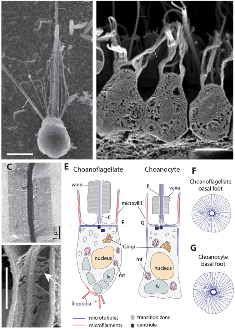
Over 600 million years ago, animals evolved from a unicellular or colonial organism whose cell(s) captured bacteria with a collar complex, a flagellum surrounded by a microvillar collar. Using principles from evolutionary cell biology, we reason that the transition to multicellularity required modification of pre-existing mechanisms for extracellular matrix synthesis and cytokinesis. We discuss two hypotheses for the origin of animal cell types: division of labor from ancient plurifunctional cells and conversion of temporally alternating phenotypes into spatially juxtaposed cell types. Mechanistic studies in diverse animals and their relatives promise to deepen our understanding of animal origins and cell biology.

Keywords: Choanozoa; choanoflagellates; evo-devo; evolutionary cell biology; metazoan origins; multicellularity.


Figures

Figure 1.
Figure 1.. Phylogenetic distribution of traits inferred in the Urmetazoan.
The presence of epithelia (Leys et al. 2009), sperm, eggs and multicellularity (Nielsen 2012) and collar complex (see Supplementary Table 1 for details and references) are mapped onto a consensus eukaryotic phylogeny modified from (Struck et al. 2014; Borner et al. 2014; Laumer et al. 2015; Torruella et al. 2015; Telford et al. 2015; Cannon et al. 2016). The collar complex is inferred to have been present in the Urchoanozoan, and to be a choanozoan synapomorphy. The relationships among sponges (Porifera), ctenophores and other animals are depicted as a polytomy to reflect uncertainties regarding their order of divergence (King and Rokas 2017). Species silhouettes are from PhyloPic (http://phylopic.org).
Figure 2.
Figure 2.. Conserved morphology and ultrastructure of choanoflagellates and sponge choanocytes.
(A, B) The collar complex is conserved in choanoflagellates and sponge collar cells. Both choanoflagellates (A, S. rosetta from (Dayel et al. 2011)) and sponge choanocytes (B, Sycon coactum, from (Leys and Hill 2012)) possess a flagellum (fl), microvilli (mv), a nucleus (nu) and a food vacuole (fv) in the same overall orientation. (C,D) A flagellar vane is present in both choanoflagllates (C, Salpingoeca amphoridium, from (Leadbeater 2014)) and choanocytes (D, Spongilla lacustris, from (Mah et al. 2014); arrow shows the fibrous structure of the vane and lateral contact with the collar). (E) Comparative ultrastructural schematics of a choanoflagellate and a sponge choanocyte, modified from (Maldonado 2004) following (Woollacott and Pinto 1995) for the microtubule cytoskeleton and (Karpov and Leadbeater 1998) for the actin cytoskeleton. (Although filopodia may occasionally be present in choanocytes, as reported in sketches from earlier studies of calcareous sponges (notably Sycon raphanus) (Duboscq and Tuzet 1939; Grassé 1973) and in one scanning electron microscopy study of the demosponge Ephydatia fluviatilis (Weissenfels 1982), they have not been reported so far in transmission electron microscopy or immunofluorescence studies and are thus not indicated here.) (mt): mitochondria. (F,G) basal microtubular foot supporting the flagellum in choanoflagellates and choanocytes, following (Garrone 1969; Woollacott and Pinto 1995; Leadbeater 2014).
Figure 3.
Figure 3.. Clonal and aggregative multicellularity.
(A) Phylogenetic distribution of clonal and aggregative multicellularity. Eukaryotic phylogeny is modified from (Keeling et al. 2014). Instances of multicellularity are mapped following (Bonner 1998; King 2004; Raven 2005; Ott et al. 2015; Sebé-Pedrós et al. 2017). (B and C) Examples of aggregative and clonal multicellularity. The organism indicated is shown in bold, and other organisms with similar forms of multicellularity are listed below. (B) Aggregative multicellularity gives rise to spherical masses of spores or cysts, sometimes atop a stalk. From (Brown et al. 2012; Du et al. 2015). (C) Clonal multicellularity gives rise to diverse multicellular forms. From (Bonner 1998; Fairclough et al. 2010).
Figure 4.
Figure 4.. Morphogenesis in choanoflagellate rosettes, calcareous sponge embryos and volvocale embryos.
(A) Morphogenesis during rosette formation in the choanoflagellate S. rosetta, following (Fairclough et al. 2010). (B) Early embryonic development of the calcareous sponge Sycon ciliatum, including amphiblastula inversion, from (Franzen 1988). (C) Early embryonic development of the volvocale Pleodorina californica (Höhn and Hallmann 2016). In other volvocales such as Volvox, an additional developmental stage is intercalated, in which the embryo first forms a sphere with flagella pointing inward, which later opens up into a continuously bending sheet that finally closes into a sphere with flagella pointing outward.
Figure 5.
Figure 5.. Temporally alternating cell types in protozoans.
(A) The heterolobosean excavate Naegleria gruberi can switch between a flagellated swimmer phenotype and a deformable crawler (“amoeboid”) phenotype. Redrawn from (Fritz-Laylin et al. 2010). (B) Cell types and life history transitions in the choanoflagellate S. rosetta, from (Dayel et al. 2011; Levin and King 2013). Main panel depicts the dynamic asexual life history of S. rosetta whereas the inset indicates its sexual cycle. Dotted lines indicated inferred transitions that have not been directly observed.
Figure 6.
Figure 6.. The division of labor hypothesis.
(A) Cellular modules present in the choanoflagellate S. rosetta. On the right: choanoflagellate orthologs of the selector transcription factors that control these modules in animals, on top of a list of choanoflagellate orthologs of their animal targets (Supplementary Table 2, Supplementary Figure 3). No terminal selector is indicated for the secretion apparatus, as there seems to be no known neural terminal selector with a choanoflagellate ortholog. Dotted lines indicate that it is unknown whether the choanoflagellate transcription factors control the same genes as their animal orthologs, except for the Myc::Max complex for which regulation is indicated by computational predictions (Brown et al. 2008) and electrophoretic mobility shift assays (Young et al. 2011). (B) Cellular modules shown in panel A are segregated into distinct cell types in animals (here, the putative cell type complement of stem-eumetazoans is illustrated based on the cell types shared by cnidarians and bilaterians (Fautin and Mariscal 1991; Schmidt-Rhaesa 2007; Arendt et al. 2015)) and terminal selector transcription factors specify distinct cell types.

References

    1. Abedin M, King N (2008) The premetazoan ancestry of cadherins. Science 319:946–948. doi: 10.1126/science.1151084 - DOI - PubMed
    1. Abedin M, King N (2010) Diverse evolutionary paths to cell adhesion. Trends Cell Biol 20:734–742. doi: 10.1016/j.tcb.2010.08.002 - DOI - PMC - PubMed
    1. Achim K, Pettit J-B, Saraiva LR, et al. (2015) High-throughput spatial mapping of single-cell RNA-seq data to tissue of origin. Nat Biotechnol 33:503–509. doi: 10.1038/nbt.3209 - DOI - PubMed
    1. Adamska M (2016) Sponges as the rosetta stone of colonial-to-multicellular transition In: Niklas KJ, Newman SA (eds) Multicellularity. Origins and Evolution. MIT Press, pp 185–200
    1. Alegado RA, Brown LW, Cao S, et al. (2012) A bacterial sulfonolipid triggers multicellular development in the closest living relatives of animals. eLife 1:e00013. doi: 10.7554/eLife.00013 - DOI - PMC - PubMed

Publication types