

Feature Extraction and Classification on Esophageal X-Ray Images of Xinjiang Kazak Nationality
- PMID: 29065605
- PMCID: PMC5394892
- DOI: 10.1155/2017/4620732
Feature Extraction and Classification on Esophageal X-Ray Images of Xinjiang Kazak Nationality
Abstract
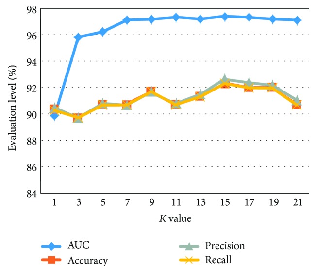
Esophageal cancer is one of the fastest rising types of cancers in China. The Kazak nationality is the highest-risk group in Xinjiang. In this work, an effective computer-aided diagnostic system is developed to assist physicians in interpreting digital X-ray image features and improving the quality of diagnosis. The modules of the proposed system include image preprocessing, feature extraction, feature selection, image classification, and performance evaluation. 300 original esophageal X-ray images were resized to a region of interest and then enhanced by the median filter and histogram equalization method. 37 features from textural, frequency, and complexity domains were extracted. Both sequential forward selection and principal component analysis methods were employed to select the discriminative features for classification. Then, support vector machine and K-nearest neighbors were applied to classify the esophageal cancer images with respect to their specific types. The classification performance was evaluated in terms of the area under the receiver operating characteristic curve, accuracy, precision, and recall, respectively. Experimental results show that the classification performance of the proposed system outperforms the conventional visual inspection approaches in terms of diagnostic quality and processing time. Therefore, the proposed computer-aided diagnostic system is promising for the diagnostics of esophageal cancer.
Figures


Similar articles
-
Differentiation of fat-poor angiomyolipoma from clear cell renal cell carcinoma in contrast-enhanced MDCT images using quantitative feature classification.Med Phys. 2017 Jul;44(7):3604-3614. doi: 10.1002/mp.12258. Epub 2017 Jun 9. Med Phys. 2017. PMID: 28376281
-
Feature extraction and pattern classification of colorectal polyps in colonoscopic imaging.Comput Med Imaging Graph. 2014 Jun;38(4):267-75. doi: 10.1016/j.compmedimag.2013.12.009. Epub 2014 Jan 2. Comput Med Imaging Graph. 2014. PMID: 24495469
-
Computer-assisted lip diagnosis on Traditional Chinese Medicine using multi-class support vector machines.BMC Complement Altern Med. 2012 Aug 16;12:127. doi: 10.1186/1472-6882-12-127. BMC Complement Altern Med. 2012. PMID: 22898352 Free PMC article.
-
Computer-aided diagnosis of pulmonary nodules on CT scans: improvement of classification performance with nodule surface features.Med Phys. 2009 Jul;36(7):3086-98. doi: 10.1118/1.3140589. Med Phys. 2009. PMID: 19673208 Free PMC article.
-
A review of image analysis and machine learning techniques for automated cervical cancer screening from pap-smear images.Comput Methods Programs Biomed. 2018 Oct;164:15-22. doi: 10.1016/j.cmpb.2018.05.034. Epub 2018 Jun 26. Comput Methods Programs Biomed. 2018. PMID: 30195423 Review.
Cited by
-
Development of a Deep Learning System to Detect Esophageal Cancer by Barium Esophagram.Front Oncol. 2022 Jun 21;12:766243. doi: 10.3389/fonc.2022.766243. eCollection 2022. Front Oncol. 2022. PMID: 35800062 Free PMC article.
-
Research status and progress of deep learning in automatic esophageal cancer detection.World J Gastrointest Oncol. 2025 May 15;17(5):104410. doi: 10.4251/wjgo.v17.i5.104410. World J Gastrointest Oncol. 2025. PMID: 40487951 Free PMC article. Review.
-
Auto-detection of cervical collagen and elastin in Mueller matrix polarimetry microscopic images using K-NN and semantic segmentation classification.Biomed Opt Express. 2021 Mar 23;12(4):2236-2249. doi: 10.1364/BOE.420079. eCollection 2021 Apr 1. Biomed Opt Express. 2021. PMID: 33996226 Free PMC article.
-
COVID-19 discrimination framework for X-ray images by considering radiomics, selective information, feature ranking, and a novel hybrid classifier.Signal Process Image Commun. 2021 Sep;97:116359. doi: 10.1016/j.image.2021.116359. Epub 2021 Jun 17. Signal Process Image Commun. 2021. PMID: 34219966 Free PMC article.
-
Detection of Collaterals from Cone-Beam CT Images in Stroke.Sensors (Basel). 2021 Dec 3;21(23):8099. doi: 10.3390/s21238099. Sensors (Basel). 2021. PMID: 34884102 Free PMC article.
References
-
- Maria A. H., Katharine A. Image-guided radiotherapy for esophageal cancer. Imaging in Medicine. 2012;4(5):515–525.
-
- Parkin D. M., Bray F. I., Devesa S. S. Cancer burden in the year 2000. The global picture. European Journal of Cancer. 2001;37(58):S4–S66. - PubMed
-
- Guang J. Y., Qian W. L., Yu X. D., et al. Analysis on the epidemiological characteristics of esophageal cancer in Huai’an area, China from 2009 to 2011. The Chinese-German Journal of Clinical Oncology. 2012;11(9):504–507. doi: 10.1007/s10330-012-1035-4. - DOI
-
- Xue Y. Z., Da F. Z., Xin M., Jiang D. Esophageal cancer spatial and correlation analyses: water pollution, mortality rates, and safe buffer distances in China. Journal of Geographical Sciences. 2014;24(1):46–58. doi: 10.1007/s11442-014-1072-8. - DOI
-
- Hui G., Jian B. D., Wei Z., Zhang T. Gene research progress on Xinjiang kazak esophageal cancer. Basic Medicine and Clinical. 2010;30(4):428–430.
Publication types
MeSH terms
LinkOut - more resources
Full Text Sources
Other Literature Sources
Medical
