Review
. 2017:2017:8427595.
doi: 10.1155/2017/8427595. Epub 2017 Sep 5.

Intraoral Scanner Technologies: A Review to Make a Successful Impression

Affiliations
Review

Intraoral Scanner Technologies: A Review to Make a Successful Impression

Raphaël Richert et al. J Healthc Eng. 2017.

Abstract

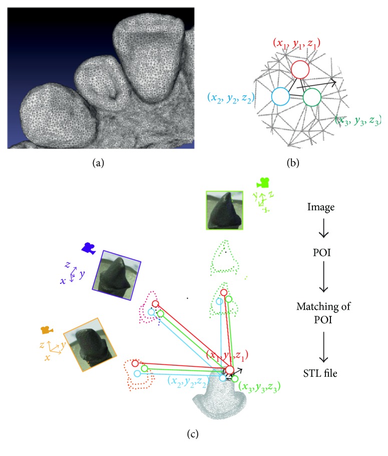
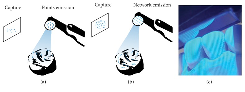
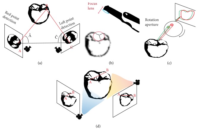
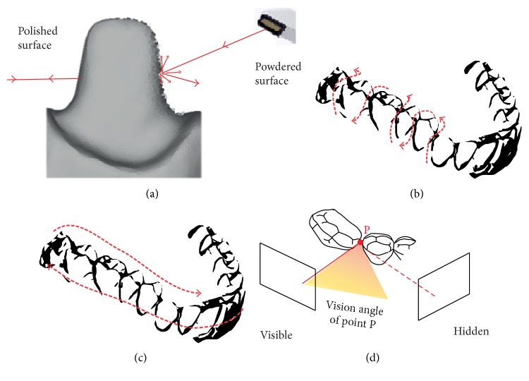
To overcome difficulties associated with conventional techniques, impressions with IOS (intraoral scanner) and CAD/CAM (computer-aided design and manufacturing) technologies were developed for dental practice. The last decade has seen an increasing number of optical IOS devices, and these are based on different technologies; the choice of which may impact on clinical use. To allow informed choice before purchasing or renewing an IOS, this article summarizes first the technologies currently used (light projection, distance object determination, and reconstruction). In the second section, the clinical considerations of each strategy such as handling, learning curve, powdering, scanning paths, tracking, and mesh quality are discussed. The last section is dedicated to the accuracy of files and of the intermaxillary relationship registered with IOS as the rendering of files in the graphical user interface is often misleading. This overview leads to the conclusion that the current IOS is adapted for a common practice, although differences exist between the technologies employed. An important aspect highlighted in this review is the reduction in the volume of hardware which has led to an increase in the importance of software-based technologies.


Figures

Figure 1
Figure 1
Generation of a STL file by intraoral scanner. (a) An example of a STL file. (b) Each triangle of a STL file is composed by three points with cartesian coordinates (x, y, and z) and a normal surface. (c) Schematic representation of the reconstruction technology: each picture is analyzed, and POI (points of interest) are selected by the software. After similarity calculation between different images, a matching of coinciding POI is defined and triangles with coordinates are generated by projection matrix.
Figure 2
Figure 2
Nature of light. (a) Projection of points. (b) Projection of a mesh. (c) Projection of a mesh by an intraoral scanner.
Figure 3
Figure 3
Determining distance to the object. (a) Triangulation: distance BC could be determined according to the formula BC=AC×sinA^/sinA^+C^. (b) Confocal: distance to the object is determined according to the focal distance. (c) AWS requiring a camera and an off-axis that moves on a circular path around the optical axis and produces a rotation of interest points. (d) Stereophotogrammetry is a technology that generates files by algorithm analyzing numerous pictures.
Figure 4
Figure 4
Scanning strategies. (a) Prepared teeth have reflective surfaces due to enamel or polished surface. Powdering can increase diffuse light that diminish this phenomenon. (b) A one-way scan (S sweep on vestibular, occlusal, and lingual surfaces). (c) A linear movement on occlusal-palatal surfaces followed by buccal surface. (d) Proximal faces are hidden if the scanning strategy is not adapted.
Figure 5
Figure 5
Management of mesh quality. Comparison of STL files depending on mesh density. (a) Low density. (b) Medium density. (c) High density. (d) Large number of triangles over the whole tooth. (e) Routine mesh on flat zones and denser mesh for gingival sulcus. (f) Prepared teeth present various points that are complex to scan. (g) Complex points can appear smoothed on CAD-CAM software. (h) Saliva or water film can generate errors during margin impression that could reduce mesh quality.
Figure 6
Figure 6
Accuracy of full arch impression and matching process. (a) Full-arch files generated with IOS and laboratory scanner were matched (with Geomagic software). (b) Three-dimensional deviations between IOS and reference files revealed posterior distortion. Impact of selected points (1, 2, and 3) to the matching process (with CloudCompare). (c) Anterior points. (d) Lateral-located points. (e) Scattered points.

References

    1. Chen L. C., Xu Z. Q. Innovative 3D dental measurement for tooth model restoration. Key Engineering Materials. 2005;295–296(12):145–150.
    1. Hong-Seok P., Chintal S. Development of high speed and high accuracy 3D dental intra oral scanner. Procedia Engineering. 2015;100:1174–1181. doi: 10.1016/j.proeng.2015.01.481. - DOI
    1. Ali A. O. Accuracy of digital impressions achieved from five different digital impression systems. Dentistry. 2015;5:p. 5.
    1. Duret F. Toward a new symbolism in the fabrication of prosthetic design. Les Cahiers de Prothèse. 1985;13(50):65–71. - PubMed
    1. Baheti M. J., Soni U. N., Gharat N. V., Mahagaonkar P., Khokhani R., Dash S. Intra-oral scanners: a new eye in dentistry. Austin Journal of Orthopedics & Rheumatology. 2015;2(3)

Substances