. 2017:2017:9547896.
doi: 10.1155/2017/9547896. Epub 2017 Aug 20.

Gentamicin Released from Porous Scaffolds Fabricated by Stereolithography

Affiliations

Gentamicin Released from Porous Scaffolds Fabricated by Stereolithography

Somruethai Channasanon et al. J Healthc Eng. 2017.

Abstract

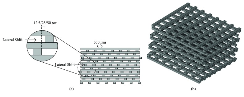
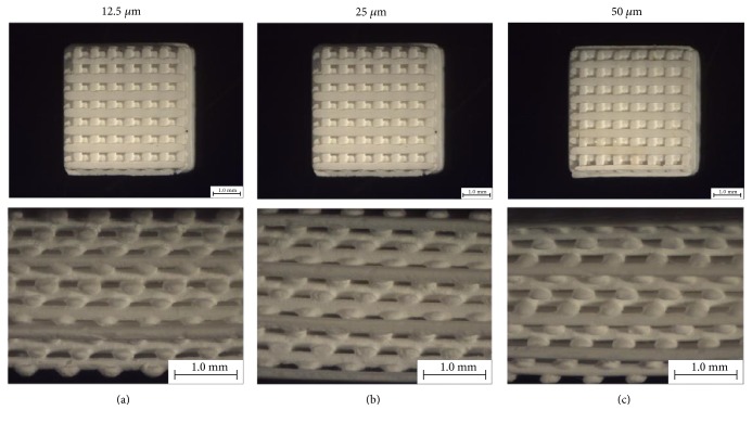
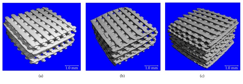
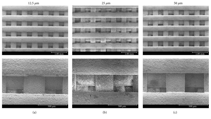
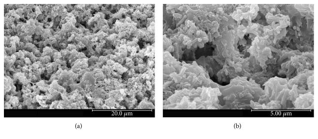
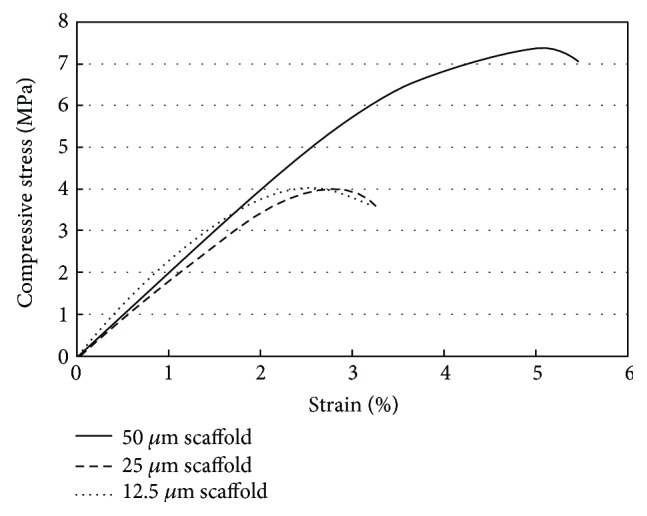
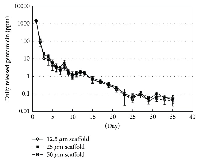
Porous oligolactide-hydroxyapatite composite scaffolds were obtained by stereolithographic fabrication. Gentamicin was then coated on the scaffolds afterwards, to achieve antimicrobial delivery ability to treat bone infection. The scaffolds examined by stereomicroscope, SEM, and μCT-scan showed a well-ordered pore structure with uniform pore distribution and pore interconnectivity. The physical and mechanical properties of the scaffolds were investigated. It was shown that not only porosity but also scaffold structure played a critical role in governing the strength of scaffolds. A good scaffold design could create proper orientation of pores in a way to strengthen the scaffold structure. The drug delivery profile of the porous scaffolds was also analyzed using microbiological assay. The release rates of gentamicin from the scaffolds showed prolonged drug release at the levels higher than the minimum inhibitory concentrations for S. aureus and E. coli over a 2-week period. It indicated a potential of the scaffolds to serve as local antibiotic delivery to prevent bacterial infection.


Figures

Figure 1
Figure 1
A computer-aided design (CAD) model of the woodpile scaffold: cross-section (a) and 3D structure (b).
Figure 2
Figure 2
Top view at 10x magnification (top) and side view at 25x magnification (bottom) of the scaffolds observed under stereomicroscope: 12.5 μm scaffold (a), 25 μm scaffold (b), and 50 μm scaffold (c).
Figure 3
Figure 3
3D images of the scaffolds by microcomputed tomography (μCT): 12.5 μm scaffold (a), 25 μm scaffold (b), and (c) 50 μm scaffold (c).
Figure 4
Figure 4
SEM micrographs at 30x magnification (top) and 110x magnification (bottom) of the scaffolds: 12.5 μm scaffold (a), 25 μm scaffold (b), and 50 μm scaffold (c).
Figure 5
Figure 5
Micropores at the scaffold surface observed by SEM: 2,500x magnification (a) and 10,000x magnification (b).
Figure 6
Figure 6
Comparison of mechanical curves of 12.5 μm, 25 μm, and 50 μm scaffolds.
Figure 7
Figure 7
Daily released gentamicin from various scaffolds incubated in PBS at 37°C: 12.5 μm scaffold (◊), 25 μm scaffold (■), and 50 μm scaffold (○).

Similar articles

Cited by

References

    1. Chen V. J., Smith L. A., Ma P. X. Bone regeneration on computer-designed nano-fibrous scaffolds. Biomaterials. 2006;27:3973–3979. doi: 10.1016/j.biomaterials.2006.02.043. - DOI - PubMed
    1. Vaquette C., Frochot C., Rahouadj R., Wang X. An innovative method to obtain porous PLLA scaffolds with highly spherical and interconnected pores. Journal of Biomedical Materials Research Part B: Applied Biomaterials. 2008;86(1):9–17. doi: 10.1002/jbm.b.30982. - DOI - PubMed
    1. Grinberg O., Binderman I., Bahar H., Zilberman M. Highly porous bioresorbable scaffolds with controlled release of bioactive agents for tissue-regeneration applications. Acta Biomaterialia. 2010;6:1278–1287. doi: 10.1016/j.actbio.2009.10.047. - DOI - PubMed
    1. Rowlands A. S., Lim S. A., Martin D., Cooper-White J. J. Polyurethane/poly(lactic-co-glycolic) acid composite scaffolds fabricated by thermally induced phase separation. Biomaterials. 2007;28:2109–2121. doi: 10.1016/j.biomaterials.2006.12.032. - DOI - PubMed
    1. Montjovent M. O., Mathieu L., Schmoekel H., et al. Repair of critical size defects in the rat cranium using ceramic-reinforced PLA scaffolds obtained by supercritical gas foaming. Journal of Biomedical Materials Research, Part A. 2007;83(1):41–51. doi: 10.1002/jbm.a.31208. - DOI - PubMed

Publication types

LinkOut - more resources