Case Reports
. 2017 Oct 23:2017:bcr2017220722.
doi: 10.1136/bcr-2017-220722.

Giant mediastinal parathyroid adenoma presenting as bilateral brown tumour of mandible: a rare presentation of primary hyperparathyroidism

Affiliations
Case Reports

Giant mediastinal parathyroid adenoma presenting as bilateral brown tumour of mandible: a rare presentation of primary hyperparathyroidism

Shibojit Talukder et al. BMJ Case Rep. .

Abstract

Hyperparathyroidism (HPT) is becoming increasingly common endocrinopathy in clinical practice. Nowadays, it is mostly diagnosed in subclinical or early clinical stage. Bony involvement in HPT has seen significant fall in incidence. Brown tumour of bone is exceptionally rare as a first manifestation of primary HPT (PHPT). Its radiological and histopathological features may be mistaken for other bony pathologies. If possibility of underlying HPT is overlooked the disease is bound to recur after surgery adding to morbidity of the patient. Here we present a case of bilateral brown tumour of mandible which was mistakenly treated as giant cell granuloma by surgical curettage. That the patient was harbouring an ectopic parathyroid adenoma with hypercalcemia causing non-specific symptoms was missed by the referring physician. This led to recurrence of the lesion. On subsequent evaluation, a giant mediastinal parathyroid adenoma causing PHPT was detected at our centre and was removed via mini sternotomy approach.

Keywords: calcium and bone; oral and maxillofacial surgery.


Conflict of interest statement

Competing interests: None declared.

Figures

Figure 1
Figure 1
Orthopantomogram (OPG) showing bilateral lytic lesions in the body of mandible with cortical thinning and medullary lucency.
Figure 2
Figure 2
Contrast enhanced MRI (CEMRI) axial image showing poorly encapsulated soft tissue lytic lesion in both half of mandible.
Figure 3
Figure 3
(A) Photomicrograph mandibular curettage specimen showing the lesion with scattered spatially distributed multinucleate giant cells with few reactive woven bony trabeculae in the left side of the photo and the native (lamellar) bony trabeculae in the right side (H&E; 40×). (B) The number of nuclei in the giant cells was much less than that of the giant cells of giant cell tumour (H&E; 400×). (C) The background spindle cells were arranged in sheets with short fascicles and vague whorling pattern (H&E; 100×); (D) The nuclear morphology of the giant cells and stromal spindle-shaped cells was different (H&E; 400×).
Figure 4
Figure 4
Whole body Tc99m sestamibi scan image showing tracer avid mediastinal lesion.
Figure 5
Figure 5
(A) Axial CT image showing superior mediastinal lesion abutting manubrium sterni (white arrow). (B) Axial view and (C) coronal view of choline positron emission CT (choline PECT) (maximum intensity projection) of the same showing intense tracer uptake in the lesion (white arrows). (D) Increased tracer uptake is noted in both half of the body of mandible (white arrow).
Figure 6
Figure 6
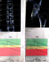
Dual-energy X-ray absorptiometry (DEXA) scan images of lumbar spine (A) and hip joint (B) showing osteopenic changes with reduced T-scores.
Figure 7
Figure 7
(A) Intraoperative photo of the giant parathyroid adenoma (GPTA) (Ad) in the superior mediastinum. Its relation to thyroid gland (Th), trachea (Tr) and right common carotid artery (C) is shown. (B) The well-encapsulated gland (Ad) is being dissected out from mediastinal fat.
Figure 8
Figure 8
(A) The microphotograph of the resected tumour showing a well-circumscribed tumour with peripherally compressed normal parathyroid gland (upper part) (H&E; 40×). The tumour showed nesting (B) and trabecular (C) patterns (H&E; 100× (B) and 200× (C)). (D) The tumour cells were relatively monomorphic and had centrally placed round nuclei, stippled chromatin, inconspicuous nuclei and moderate amount of eosinophilic to clear cytoplasm (H&E; 400×).
Figure 9
Figure 9
One-year follow-up orthopantomogram (OPG) showing healing of both the brown tumours with sclerosis.

References

    1. Parbatani R, Tinsley GF, Danford MH. Primary hyperparathyroidism presenting as a giant-cell epulis. Oral Surg Oral Med Oral Pathol Oral Radiol Endod 1998;85:282–4. 10.1016/S1079-2104(98)90009-9 - DOI - PubMed
    1. Kumar S, Bhate K, Pawar V, et al. . Osteitis fibrosa cystica of jaws as diagnostic criteria for hyperparathyroidism–a case report with review of literature. Int J Med Pharm Case Reports 2016;6:1–6. 10.9734/IJMPCR/2016/24344 - DOI
    1. Garas G, Poulasouchidou M, Dimoulas A, et al. . Radiological considerations and surgical planning in the treatment of giant parathyroid adenomas. Ann R Coll Surg Engl 2015;97:e64–e66. 10.1308/003588415X14181254789682 - DOI - PMC - PubMed
    1. Wanzari PV, Chaudhary A, Reddy V, et al. . Oral manifestations established the diagnosis of hyperparathyroidism: a rare case report. Journal of Indian Academy of Oral Medicine and Radiology 2011;23:155–8. 10.5005/jp-journals-10011-1118 - DOI
    1. Neagoe RM, Sala DT, Borda A, et al. . Clinicopathologic and therapeutic aspects of giant parathyroid adenomas - three case reports and short review of the literature. Rom J Morphol Embryol 2014;55:669–74. - PubMed

Publication types

MeSH terms

Substances