Case Reports
. 2018 Apr-Jun;64(2):115-118.
doi: 10.4103/jpgm.JPGM_739_16.

Rare tracheal tumor: Solitary plasmacytoma

Affiliations
Case Reports

Rare tracheal tumor: Solitary plasmacytoma

R Stevic et al. J Postgrad Med. 2018 Apr-Jun.

Abstract

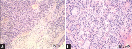
Primary tracheal tumors are rare and trachea is an exceedingly rare site of extramedullary plasmacytoma (EMP). We report a case of solitary tracheal plasmacytoma causing symptoms of airway obstruction in a 59-year-old man. Flow/volume loop indicated the fixed central airway obstruction. Computerized tomography and bronchoscopy demonstrated a sessile tumor on posterior tracheal wall obstructing 80% of the lumen. Partial tracheal resection with T-T anastomosis was performed. Pathologic analysis of resected mass revealed EMP. Additional investigations excluded multiple myeloma. There are no signs of disease recurrence after 7-year follow-up.

Keywords: Extramedullary plasmacytoma; solitary; trachea.


Conflict of interest statement

There are no conflicts of interest

Figures

Figure 1
Figure 1
Axial computerized tomography scan (a) shows an intraluminal mass, 15-mm in diameter, arising from the membranous part of the middle-third of the trachea and obstructing the lumen. Coronal (b) and sagittal (c) reformation showed the longitudinal extent of tumor; upper part is 3.5 cm distal to vocal cords (blue line)
Figure 2
Figure 2
Virtual bronchoscopy shows spherical, smooth-surfaced tumor obstructing lumen of trachea (arrows)
Figure 3
Figure 3
Morphology: Monotonic cell population proliferation was detected in outer layer of tracheal wall (a); on higher magnification well-differentiated plasmacytic proliferation was seen (b)
Figure 4
Figure 4
Immunohistochemistry: Plasma cell origin of proliferation was confirmed by CD38 (a) and CD138 (b); only kappa light chain immunoglobulin expression was found in plasma cells (c); lambda light chain immunoglobulin expression was not found, and monoclonality of plasma cells was proven (d)

References

    1. Kober SJ. Solitary plasmacytoma of the carina. Thorax. 1979;34:567–8. - PMC - PubMed
    1. Garelli M, Righini C, Faure C, Jankowski A, Brambilla C, Ferretti GR. Imaging of a case of extramedullary solitary plasmacytoma of the trachea. Case Rep Radiol. 2011;2011:687203. - PMC - PubMed
    1. Kairalla RA, Carvalho CR, Parada AA, Alves VA, Saldiva PH. Solitary plasmacytoma of the trachea treated by loop resection and laser therapy. Thorax. 1988;43:1011–2. - PMC - PubMed
    1. Sukumaran R, Nair RA, Jacob PM, Koshy SM, Mathew AP. Extramedullary plasmacytoma of the trachea. Head Neck Pathol. 2014;8:220–4. - PMC - PubMed
    1. Cuttitta A, Tancredi A, Scaramuzzi R, Falcone A, Scaramuzzi G, Taurchini M. Solitary extramedullary plasmacytoma of the trachea: A case report. Int J Cardiovasc Thorac Surg. 2016;2:5–8.

Publication types