. 2017 Oct 25;9(11):331.
doi: 10.3390/toxins9110331.

Soluble CD40 Ligand and Oxidative Response Are Reciprocally Stimulated during Shiga Toxin-Associated Hemolytic Uremic Syndrome

Affiliations

Soluble CD40 Ligand and Oxidative Response Are Reciprocally Stimulated during Shiga Toxin-Associated Hemolytic Uremic Syndrome

Maria J Abrey Recalde et al. Toxins (Basel). .

Abstract

Shiga toxin (Stx), produced by Escherichia coli, is the main pathogenic factor of diarrhea-associated hemolytic uremic syndrome (HUS), which is characterized by the obstruction of renal microvasculature by platelet-fibrin thrombi. It is well known that the oxidative imbalance generated by Stx induces platelet activation, contributing to thrombus formation. Moreover, activated platelets release soluble CD40 ligand (sCD40L), which in turn contributes to oxidative imbalance, triggering the release of reactive oxidative species (ROS) on various cellular types. The aim of this work was to determine if the interaction between the oxidative response and platelet-derived sCD40L, as consequence of Stx-induced endothelium damage, participates in the pathogenic mechanism during HUS. Activated human glomerular endothelial cells (HGEC) by Stx2 induced platelets to adhere to them. Although platelet adhesion did not contribute to endothelial damage, high levels of sCD40L were released to the medium. The release of sCD40L by activated platelets was inhibited by antioxidant treatment. Furthermore, we found increased levels of sCD40L in plasma from HUS patients, which were also able to trigger the respiratory burst in monocytes in a sCD40L-dependent manner. Thus, we concluded that platelet-derived sCD40L and the oxidative response are reciprocally stimulated during Stx2-associated HUS. This process may contribute to the evolution of glomerular occlusion and the microangiopathic lesions.

Keywords: CD40L; Shiga toxin 2; blood platelets; hemolytic uremic syndrome; oxidative stress.


Conflict of interest statement

The authors declare that they have no conflict of interest.

Figures

Figure 1
Figure 1
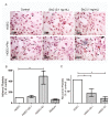
Microvasculature damage by Stx2. Human glomerular endothelial cells (HGEC) were seeded on gelatin-coated glass coverslips (A), or 24-well plates (B,C) treated or not with 0.1 ng/mL (S0.1), 1 ng/mL (S1) Stx2, 1 ng/mL Stx2 inactivated by heating (S1 inact) or 1 ng/mL Stx2B (Stx2B). After 24 h, platelets (Plts) (1 × 10⁸/well) were added at 37 °C in 5% CO2 for 1 h and washed. (A) Representative images by light microscopy from each experimental condition stained with H&E (×10) are shown. Viability was evaluated 1 h (B) or 18 h (C) after Plts addition. Cells were incubated with neutral red an additional hour at 37 °C in 5% CO2. The percentage of viable cells was calculated considering that absorbance (A) obtained for cells incubated without toxin treatment represents 100% viability. Each condition was made in duplicate for each experiment. Data are expressed as median and interquartile range of three independent experiments (n = 3). * p < 0.05.
Figure 2
Figure 2
Platelet adhesion to damaged endothelium. HGEC were seeded on gelatin-coated glass coverslips (A), or 24-well plates (B,C) treated or not with 0.1 ng/mL (S0.1) or 1 ng/mL (S1) Stx2. After 24 h, Plts (1 × 10⁸/well) were added for 1 h at 37 °C in 5% CO2 and washed. (A) Representative images by light microscopy from each experimental condition stained with H&E (×400) are shown. HGEC are indicated by asterisk and Platelets by arrows. (B) Cells were incubated with PNP and acid phosphatase activity was measured reading A at 405 nm. A obtained in wells with HGEC without platelets was subtracted from that obtained in wells with platelets for each experimental condition. The percentage of adhered platelets was calculated considering that wells without Stx2 treatment represents 100%. The control of platelet adhesion to gelatin-covered well is shown as medium. (C) Supernatants from HGEC-Plts cultures were collected and the number of Plts recovered was determined by hematology analyzer. The number of Plts recovered in HGEC cultures without Stx2 treatment represented 100%. Each condition was made in duplicate for each experiment. Data are expressed as median and interquartile range of five independent experiments (n = 5). * p < 0.05.
Figure 3
Figure 3
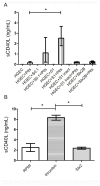
Release of sCD40L by Plts. (A) HGEC were placed in gelatin-covered 24-well plates treated or not with 0.1 ng/mL (S0.1), 1 ng/mL (S1) Stx2, 1 ng/mL Stx2 inactivated by heating (S1 inact) or 1 ng/mL Stx2B. After 24 h, Plts (1 × 10⁸/well) were added for 1 h at 37 °C in 5% CO2 and washed. Supernatants of HGEC cultures were collected and sCD40L levels were measured by ELISA kit. (B) Isolated Plts (1 × 10⁸/well) were stimulated with 0.2 U/mL thrombin or 1 ng/mL Stx2 during 1 h at 37 °C in 5% CO2 and centrifuged. Supernatants were collected and sCD40L levels measured by ELISA kit. Each condition was made in duplicate for each experiment. Data are expressed as median and interquartile range of eight independent experiments (n = 8). * p < 0.05. Controls with S1 inact and Stx2B were performed 3 times and each condition was made by duplicate.
Figure 4
Figure 4
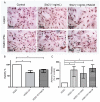
Role of oxidative stress in endothelial damage and platelet adhesion. HGEC were seeded on gelatin-covered glass coverslips (A) or 24-well plates (B,C) treated or not with 1 ng/mL (S1) Stx2. After 24 h, Plts (1 × 10⁸/well) were added for 1 h at 37 °C in 5% CO2. NAC (1 nM) was incorporated simultaneously with Stx2 (NAC#) or previous to Plts (NAC). (A) Representative images by light microscopy from each experimental condition stained with H&E (×400). Number of viable cells and Plts adhered to HGEC were observed by light microscopy (×400). HGEC are indicated by asterisk and Platelets by arrows; (B) Cells were incubated with neutral red for an additional 1 h at 37 °C in 5% CO2. The percentage of viable cells was calculated considering that A obtained for cells incubated without toxin treatment represents 100% viability; (C) Cells were incubated with PNP and acid phosphatase activity was measured reading A at 405 nm. A obtained in wells with HGEC without platelets was subtracted from that obtained in wells with platelets. The percentage of adhered platelets was calculated considering that wells without Stx2 treatment represents 100% of adhered platelets. Each condition was made in duplicate for each experiment. Data are expressed as median and interquartile range of eight independent experiments (n = 8). * p < 0.05.
Figure 5
Figure 5
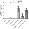
Role of oxidative stress in sCD40L platelet release. HGEC were placed in 24-well plates treated or not with 1 ng/mL (S1) Stx2. After 24 h Plts (1 × 10⁸/well) were added for 1 h at 37 °C in 5% CO2. NAC (1 nM) was incorporated simultaneously with Stx2 (NAC#) or previous to Plts (NAC). Supernatants of HGEC cultures were collected and sCD40L levels were measured by ELISA kit. Each condition was made in duplicate for each experiment. Data are expressed as median and interquartile range of eight independent experiments (n = 8). * p < 0.05, *** p< 0.001.
Figure 6
Figure 6
sCD40L levels in hemolytic uremic syndrome (HUS) plasma. (A) sCD40L levels in HUS patients and healthy controls (HC) plasmas were determined by ELISA kit. In addition, HUS patients were retrospectively classified according to Gianantonio et al.’s criteria in grade 1/2 (n = 13) and grade 3 (n = 10). Data are expressed as median and interquartile range; (B) Quantities of sCD40L (ng) released per 1 × 10⁸ Plts from HUS patients and HC. Data are expressed as median and interquartile range; (C) Correlation between sCD40L and plama creatinine levels in HUS patients; (D) Correlation between sCD40L and plasma urea levels in HUS patients. Points represent independent individuals. * p < 0.05 *** p < 0.001.
Figure 7
Figure 7
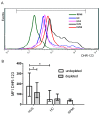
Reactive oxidative species (ROS) production by monocytes. Peripheral blood mononuclear cells (PBMC) were incubated 1h with plasmas from HUS patients or HC. Then, PMBC were washed and incubated with DHR-123 and ROS production by monocytes was measured by flow cytometry. Monocytic population was gated by FSC-H/SSC-H and CD14 staining. (A) Representative histogram of one experiment: PBMC incubated with: HC (healthy control plasma), HCd (Healthy control plasma depleted of sCD40L), HUS (HUS plasma) and HUSd (HUS plasma depleted of sCD40L; (B) Each bar shows the mean fluorescence intensity (MFI) of monocytes from 6 independent donors under different conditions. Data are expressed as median and interquartile range. * p < 0.05.

Similar articles

Cited by

References

    1. Palermo M.S., Exeni R.A., Fernandez G.C. Hemolytic uremic syndrome: Pathogenesis and update of interventions. Expert Rev. Anti-Infect. Ther. 2009;7:697–707. doi: 10.1586/eri.09.49. - DOI - PubMed
    1. Schuller S., Frankel G., Phillips A.D. Interaction of shiga toxin from Escherichia coli with human intestinal epithelial cell lines and explants: Stx2 induces epithelial damage in organ culture. Cell. Microbiol. 2004;6:289–301. doi: 10.1046/j.1462-5822.2004.00370.x. - DOI - PubMed
    1. O’Brien A.D., Tesh V.L., Donohue-Rolfe A., Jackson M.P., Olsnes S., Sandvig K., Lindberg A.A., Keusch G.T. Shiga toxin: Biochemistry, genetics, mode of action, and role in pathogenesis. Curr. Top. Microbiol. Immunol. 1992;180:65–94. - PubMed
    1. Gallo E.G., Gianantonio C.A. Extrarenal involvement in diarrhoea-associated haemolytic-uraemic syndrome. Pediatr. Nephrol. 1995;9:117–119. doi: 10.1007/BF00858990. - DOI - PubMed
    1. Ruggenenti P., Noris M., Remuzzi G. Thrombotic microangiopathy, hemolytic uremic syndrome, and thrombotic thrombocytopenic purpura. Kidney Int. 2001;60:831–846. doi: 10.1046/j.1523-1755.2001.060003831.x. - DOI - PubMed

Publication types

MeSH terms

LinkOut - more resources