. 2018 Jan;39(1):152-166.
doi: 10.1002/humu.23362. Epub 2017 Nov 6.

Expanding the genetic architecture and phenotypic spectrum in the skeletal ciliopathies

Affiliations

Expanding the genetic architecture and phenotypic spectrum in the skeletal ciliopathies

Wenjuan Zhang et al. Hum Mutat. 2018 Jan.

Abstract

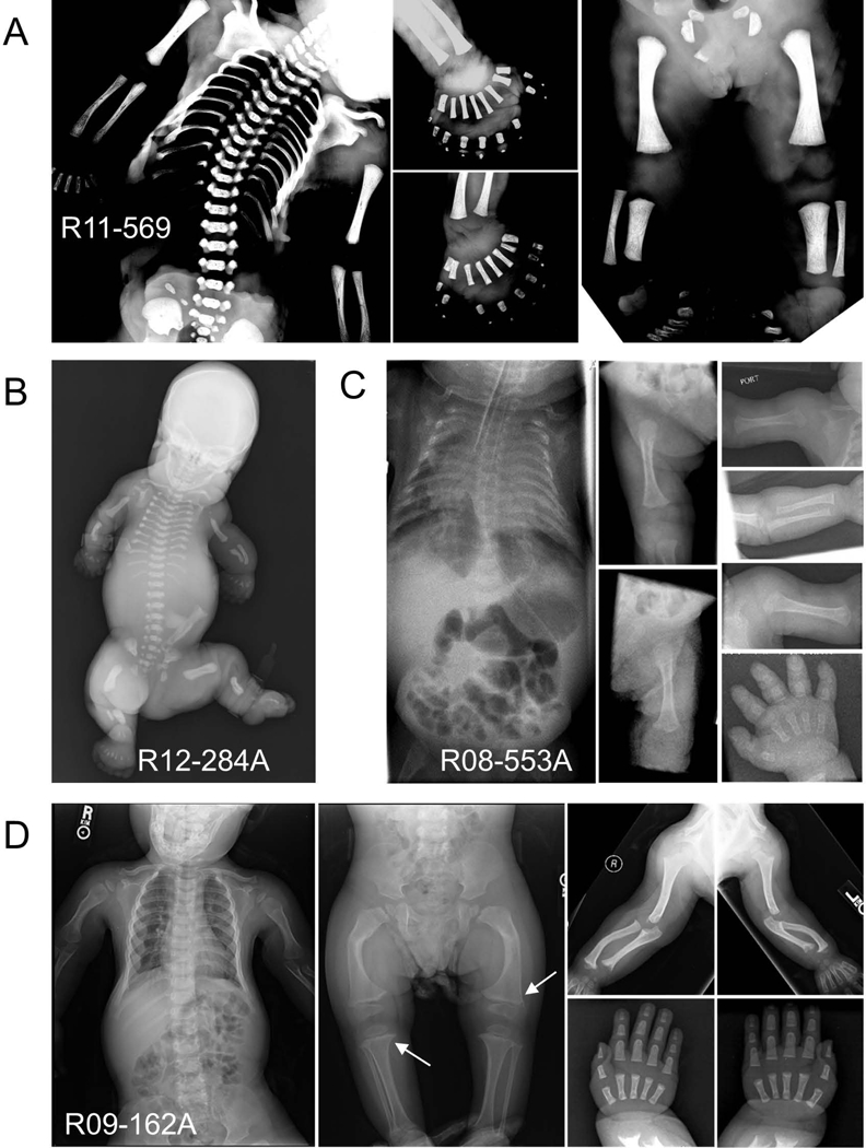
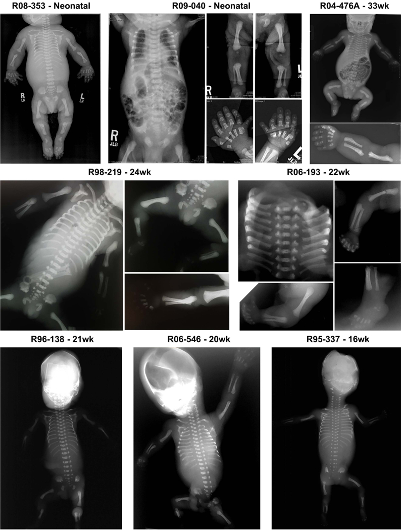
Defects in the biosynthesis and/or function of primary cilia cause a spectrum of disorders collectively referred to as ciliopathies. A subset of these disorders is distinguished by profound abnormalities of the skeleton that include a long narrow chest with markedly short ribs, extremely short limbs, and polydactyly. These include the perinatal lethal short-rib polydactyly syndromes (SRPS) and the less severe asphyxiating thoracic dystrophy (ATD), Ellis-van Creveld (EVC) syndrome, and cranioectodermal dysplasia (CED) phenotypes. To identify new genes and define the spectrum of mutations in the skeletal ciliopathies, we analyzed 152 unrelated families with SRPS, ATD, and EVC. Causal variants were discovered in 14 genes in 120 families, including one newly associated gene and two genes previously associated with other ciliopathies. These three genes encode components of three different ciliary complexes; FUZ, which encodes a planar cell polarity complex molecule; TRAF3IP1, which encodes an anterograde ciliary transport protein; and LBR, which encodes a nuclear membrane protein with sterol reductase activity. The results established the molecular basis of SRPS type IV, in which mutations were identified in four different ciliary genes. The data provide systematic insight regarding the genotypes associated with a large cohort of these genetically heterogeneous phenotypes and identified new ciliary components required for normal skeletal development.

Keywords: chondrocyte; cilia; ciliopathy; skeletal dysplasia.


Figures

FIGURE 1
FIGURE 1
Radiographic phenotypes of the SRPS probands with mutations in FUZ, TRAF3IP1 and LBR. (a) FUZ mutations in SRPS type II (R11–569, 24 weeks) showing a long, narrow chest, moderately short ribs, short long bones, hypoplastic tibiae and extreme polydactyly of all four limbs. (b) TRAF3IP1 mutations in SRPS type II (R12–284A, neonatal) showing short long bones, very short horizontal ribs and long narrow chest, severe micromelia and polydactyly of all limbs of the affected individuals. (c) TRAF3IP1 mutations in ATD (R08–553A, neonatal) showing a bell-shaped thorax, moderately short ribs, handlebar clavicles, micromelia of all long bones and brachydactyly. (d) LBR mutations associated with ATD (R09–162A, 2y 9m) showing moderately short horizontal ribs, short limbs, metaphyseal abnormalities (white arrows) and no polydactyly.
FIGURE 2
FIGURE 2
Mutations in DYNC2H1 in cases of ATD and SRPS. The structure of the human DYNC2H1 protein showing the locations of the 110 mutations described in this study. Font color indicates mutations associated with ATD (black), SRPS type I (fuchsia), SRPS type II (blue) and SRPS type III (red). Mutations frequencies are denoted with the number of circles and the types of mutations are distinguished with colors. The figure also includes 11 mutations from 6 previously published cases with mutations in the gene (Badiner, et al., 2016; Merrill, et al., 2009). Conserved protein domain localizations were taken from Schmidts et al., 2013 (Schmidts, et al., 2013a) with six AAA+ domains, an MT-binding stalk, an N-terminal tail (DHC_N1), a linker domain (DHC_N2) and a conserved C-terminal domain (Dynein_heavy). The plot was generated with Protein Paint.
FIGURE 3
FIGURE 3
Mutations identified in dynein motor and IFT-A proteins. The structures of the human dynein motor (WDR34 and WDR60) and IFT-A (IFT140, WDR19, WDR35 and TTC21B) proteins showing the locations of the mutations identified in this study. Mutation frequencies are denoted with the number of circles, and the mutation types are distinguished with colors as described in Figure 1. The figure includes 10 mutations from 7 previously published cases with mutations in these genes (Duran, et al., 2017; Huber, et al., 2013). Conserved protein domain regions were identified using the UniProt and InterPro websites.
FIGURE 4
FIGURE 4
Radiographic phenotype of the probands with EVC and EVC2 mutations. At later gestational ages, an EVC radiographic phenotype consisting of short long bones, moderately short horizontal ribs and a long narrow chest, a trident acetabulum, reverse campomelia of the humeri along with a distinctive configuration of the radius and ulna, polydactyly and brachydactyly with well ossified digits was evident. At earlier ages, the phenotypes more closely resembled SRPS and ATD. ISDR reference numbers and gestational ages in weeks are indicated for each individual.
FIGURE 5
FIGURE 5
Ciliary proteins mutated among the skeletal ciliopathies. The complexes in which each protein participates are shown with the mutated components identified by red asterisks.

References

    1. Adzhubei IA, Schmidt S, Peshkin L, Ramensky VE, Gerasimova A, Bork P, Kondrashov AS, Sunyaev SR. 2010. A method and server for predicting damaging missense mutations. Nature Methods 7(4):248–249. - PMC - PubMed
    1. Alby C, Piquand K, Huber C, Megarbane A, Ichkou A, Legendre M, Pelluard F, Encha-Ravazi F, Abi-Tayeh G, Bessieres B, El Chehadeh-Djebbar S, Laurent N, Faivre L, Sztriha L, Zombor M, Szabo H, Failler M, Garfa-Traore M, Bole C, Nitschke P, Nizon M, Elkhartoufi N, Clerget-Darpoux F, Munnich A, Lyonnet S, Vekemans M, Saunier S, Cormier-Daire V, Attie-Bitach T, Thomas S. 2015. Mutations in KIAA0586 Cause Lethal Ciliopathies Ranging from a Hydrolethalus Phenotype to Short-Rib Polydactyly Syndrome. American Journal of Human Genetics 97(2):311–318. - PMC - PubMed
    1. Anderson CT, Castillo AB, Brugmann SA, Helms JA, Jacobs CR, Stearns T. 2008. Primary cilia: Cellular sensors for the skeleton. Anatomical Record-Advances in Integrative Anatomy and Evolutionary Biology 291(9):1074–1078. - PMC - PubMed
    1. Arts HH, Bongers EMHF, Mans DA, van Beersum SEC, Oud MM, Bolat E, Spruijt L, Cornelissen EAM, Schuurs-Hoeijmakers JHM, de Leeuw N, Cormier-Daire V, Brunner HG, Knoers NVAM, Roepman R. 2011. C14ORF179 encoding IFT43 is mutated in Sensenbrenner syndrome. Journal of Medical Genetics 48(6):390–395. - PubMed
    1. Badano JL, Mitsuma N, Beales PL, Katsanis N. 2006. The ciliopathies: An emerging class of human genetic disorders. Annual Review of Genomics and Human Genetics 7:125–148. - PubMed