. 2017 Oct 25;12(10):e0185285.
doi: 10.1371/journal.pone.0185285. eCollection 2017.

Spatiotemporal changes in the distribution of LHFPL5 in mice cochlear hair bundles during development and in the absence of PCDH15

Affiliations

Spatiotemporal changes in the distribution of LHFPL5 in mice cochlear hair bundles during development and in the absence of PCDH15

Shanthini Mahendrasingam et al. PLoS One. .

Abstract

Mechanosensory transduction by vertebrate hair cells depends on a protein complex at the tips of shorter stereocilia associated with mechanoelectrical transduction channels activated by tip links in the hair bundle. In mammalian hair cells, this complex includes transmembrane channel-like protein subunit 1 (TMC1), lipoma HMGIC fusion partner-like 5 protein (LHFPL5) and protocadherin 15 (PCDH15), a lower-end component of the tip link. TMC1 interacts with LHFPL5 and PCDH15 but how the complex develops to maturity, and the relationships between these proteins, remains uncertain. Here we evaluate the spatiotemporal development of LHFPL5 distributions in mouse cochlear hair bundles by immunofluorescence and immunogold transmission electron microscopy, from postnatal day 0 (P0) through P21 in wild type and PCDH15-deficient mice. At P0, hair bundles contain many short microvilli-like processes which we term unranked stereocilia, and a subset of lengthening rows, adjacent to a kinocilium. LHFPL5 is distributed throughout the bundle, including on stereocilia tips and the kinocilium. At P3, 4-to-6 rows of ranked stereocilia are evident, total LHFPL5 expression peaks, and LHFPL5 is localised to ranked stereocilia tips of all rows and to lower shaft/ankle links. By P12, the bundle has a mature pattern with 3 ranked rows but virtually no unranked stereocilia or kinocilium; LHFPL5 expression has declined and become restricted to the tips of shorter stereocilia. Throughout development from P0, expression of LHFPL5 is greater overall on apical than basal bundles, but there is, on average, an equal amount of labelling per labelled tip. In P3 mice lacking PCDH15, LHFPL5 labelling is not at the tips but is primarily on unranked stereocilia and lower lateral links. These data show that LHFPL5 is already present in the MET apparatus at P0 but requires PCDH15 at P3 to remain there. Shaft/ankle link localisation suggests it interacts with link proteins other than PCDH15.


Conflict of interest statement

Competing Interests: The authors have declared that no competing interests exist.

Figures

Fig 1
Fig 1. SEM of representative stages of hair bundle development in CD/1 mouse OHCs at approximately 80% of the distance from the cochlear base.
P1 (a–d), P3 (e–g) and P7 (h–j) are illustrated. (a) At P1 bundles initially consist of large numbers of thin unranked stereocilia (u), similar in width to adjacent supporting cell microvilli, and the beginning of ranked stereocilia (r). (b) There are four to five ranked rows, with lateral links between the ranked stereocilia (inset, arrowhead). c) At higher magnification, tip links can also be seen in the ranked rows (t) and lateral links between unranked stereocilia (l). (d) The kinocilium is also linked to the tall stereocilia, most clearly at P1. (e) By P3 both ranked and unranked stereocilia become thicker with short unranked stereocilia gradually becoming reduced in number. (f) Several ranked rows (~6) become apparent behind unranked (u) stereocilia. (g) The unranked stereocilia have lateral links (l), and numerous tip links (t) are seen, sometimes more than one per tip, on the ranked rows. (h) By P7, most of the unranked (u) stereocilia and some of the short, ranked rows have gone, leaving the almost adult pattern of three ranked rows (r). (i) The lateral links have become reduced. (j) Multiple links are still seen around the tips. (k–m) The kinocilium (*) shows gradual thinning between P2 and P6, and is mostly absent by P8 at this location. Scale bars: a, e, h = 5 μm; b, f, i = 1 μm; c, d, j = 500 nm; g = 100 nm; k–m = 10 μm.
Fig 2
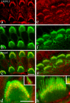
Fig 2. Confocal images of immunofluorescence labelling for LHFPL5 (red) and phalloidin (green) in CD/1 mice at approximately 80% of the distance from the cochlear base.
Upper row, P3; second row P9, and bottom row Lhfpl5-/-, P5. Both IHC and OHC hair bundles are positive for LHFPL5 at both ages. Some labelling is also detected on the apical surface, especially in CD/1 P3. The knockout shows an absence of bundle labelling. Scale bar = 20 μm.
Fig 3
Fig 3. Confocal images of immunofluorescence labelling for LHFPL5 (red) and phalloidin (green) at P9 in C3HeB/FeJ mice at approximately 80% of the distance from the cochlear base.
(a–d) distinct red fluorescence is associated with the OHC stereocilia and (d) reveals that a significant proportion of the signal lies between the stereocilia. The inset in (d) illustrates an orthogonal cut plane, indicating that most of the LHFPL5 fluorescence is in the lower part of the bundle. (e–h) Equivalent images of the IHCs show similar distribution patterns. Most of the labelling is between or on the shorter ranks and not the tallest row of stereocilia. The inset in h shows an orthogonal view similarly indicating most of the colocalised (orange) signal is in the lower part of the bundle. Scale bars: a–c, e–g = 10 μm; d, h = 5 μm.
Fig 4
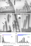
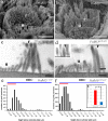
Fig 4. Semi-thin sections of pre-embedding immunogold labelling of OHC bundles from P0 to P21 at approximately 80% of the distance from the cochlear base.
(a) At P0, labelling is distributed throughout the unranked (u) and developing ranks (r) of stereocilia, and at the tips of ranked stereocilia (arrows, region enlarged 2X in inset). (b) At P3, labelling is associated with lower shaft ankle/lateral links of the stereocilia and the tips of mainly shorter and intermediate ranked stereocilia, and along the kinocilium. The kinocilium is also labelled (inset, same magnification). (c) At P12 there are three ranked rows (s = short, i = intermediate and t = tall) and very few unranked stereocilia. Labelling is confined mostly to the tips of short and intermediate ranked stereocilia. Note that some tip labelling appears displaced (see Discussion for a consideration of this effect). Apical membrane labelling is also visible (arrowhead). (d) At P21 there are still particles labelling on some tips (arrow and inset, X1.5 enlargement). (e) No labelling is detectable in the Lhfpl5-/- at P12. (f) Plot of the distribution of particles according to height from the apical membrane/cuticular plate in P3 hair bundles. The height of ranked (blue) and unranked (red) stereocilia is indicated by the horizontal lines. (At this stage, determining which of the ranked rows a stereocilium belongs to is difficult because of the minimal differences in height and the extra rows). The counts show gold particles towards the ankle/lower shafts and labelling of the apical membrane. (g) By P12, the distribution of particles is much more towards the tips (indicated by green horizontal bars) of the short and intermediate ranked rows (blue horizontal bars). Scale bars: 500 nm.
Fig 5
Fig 5
(a-e) Pre-embedding immunogold labelling of P0 to P21 IHC bundles at approximately 80% of the distance from the cochlear base. (a) At P0 some labelling is found on unranked stereocilia, but more is found over the developing ranked stereocilia and some can be seen on the kinocilium (k). (b) P3 semithin and (c) P3 ultrathin sections. Gold particles are seen along the shafts and tips at P3, including the tall stereocilia (t). The tip labelling is seen at 1.75X enlargement in the inset in c. (d) By P12 and (e) P21, only weak labelling is visible on the shafts of the stereocilia (arrowhead) but the tips of shorter rows of stereocilia are still labelled (arrows; inset X2). Scale bars = 500 nm.
Fig 6
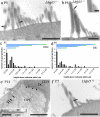
Fig 6. Postembedding immunogold labelling at approximately 80% of the distance from the cochlear base.
(a-d). Post-embedding immunogold labelling of Lhfpl5+/- at P5 confirms distributions seen between P0 and P9 in pre-embedding labelling of wild-type mice. (a) Lower shaft labelling (between arrows) and (b) tip labelling (arrow and inset) were both detected. (c, d) Counts from OHCs and IHCs revealed a tendency to label the lower parts of the bundle, but there was detectable enrichments associated with the tips (horizontal green bars) of all except the tallest stereocilia. (e) At P14 in CD/1 mice, labelling was also detected in the junctions between pillar cells (e.g. arrows) and also between pillar cells, Deiters’ cells (e.g. arrowheads) and hair cells (inset), which was not detected in pre-embedding, due probably to steric hindrance. IPC = inner pillar cell, OPC = outer pillar cell, PC = pillar cell, DC = Deiters’ cell, OHC = outer hair cell. (f) Minimal labelling was detected in Lhfpl5-/- mice–the only particle observed in 50+ sections is shown (arrow). Scale bars: a, e, e inset, f = 1 μm, b = 500 nm, b inset = 200 nm.
Fig 7
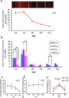
Fig 7. Quantification of immunofluorescence and immunogold labelling from P0 to P21.
a. Panel of confocal images of OHCs from the CD/1 mouse cochlea at different ages, acquired under identical conditions in the same experiment at approximately 80% of the distance from the cochlear base. Labelling was performed without phalloidin in order to eliminate the possibility of cross-talk between red and green channels in the confocal. Fluorescence intensity is high at P0 and P3 and drops after P3. No labelling was detected after P12. A graphical representation of the changes is shown below, derived from fluorescent intensity measurements of hair bundles from each of the above stages. Error bars represent the S.E.M of mean intensity measured in 10 bundles from two images. (b) Quantitative analysis of immunogold labelling in apical (at approximately 80% of the distance from the cochlear base) and basal (approximately 20–30% from the cochlear base, where possible), representing the mean of 3 different animals at each age. Counts reveal a similar high level of expression in P0 to P3 in both IHCs and OHCs, dropping substantially to a similar level over P6 –P21. Throughout development, apical labelling is higher than basal labelling, but not always significantly. Significant differences at p < 0.05, Wilcoxon (Mann-Whitney) test, are indicated by asterisks. No data could be obtained for P9 or P21 basal rgions. (c) Developmental changes in the proportion of tips that are labelled. The proportion is highest at P0 and P3 and then decreases with maturation. (d) Developmental changes in the number of particles per labelled tip. The number on each labelled tip does not change from P0 to P21. (e) Analysis of kinocilium labelling (particles per μm2) on both IHCs and OHCs reveals a peak at P3 compared with P0 and P6.
Fig 8
Fig 8. Confocal images of LHFPL5 (red) and actin/phalloidin (green) labelling in Pcdh15+/av3J and Pcdh15av3J/av3J mice at approximately 80% of the distance from the cochlear base.
(a—c) In the heterozygote, labelling appeared similar to wild type. (d—f) In the homozygote, the phalloidin revealed that bundles were distorted, and the LHFPL5 could be found over the bundle and the cuticular plate region. Scale bars = 20 μm.
Fig 9
Fig 9. SEM and TEM of labelling in Pcdh15+/av3J and Pcdh15av3J/av3J mice.
(a, b). SEM comparison of OHC hair bundles. The heterozygote is similar to wild type but the homozygote bundle is dysmorphic. Nevertheless the latter still has regions of ranked (r), as well as unranked (u) stereocilia. (c–d). Pre-embedding immunogold labelling using TEM in P3 heterozygote and homozygote hair cells at approximately 80% of the distance from the cochlear base. The heterozygote labelling resembles the wild type, but the homozygote shows predominant labelling on the unranked stereocilia. Note that lateral links occur between the unranked stereocilia, as well as the ranked stereocilia, and are still present in the homozygote (d, inset arrowhead, ultrathin section) where they are also labelled (arrow, main image). (e, f) Quantitative analysis of labelling on the heterozygote and the homozygote. Labelling is lower down the bundle in the homozygote compared with the heterozygote, and is more concentrated in the unranked stereocilia. The blue bars show the heights of the tall stereocilia, the red bar the heights of the ranked and unranked short stereocilia. (g). Relative proportion of total labelling over the unranked stereocilia in Pcdh15+/av3J and Pcdh15av3J/av3J mice OHCs. There is a significantly higher proportion on the unranked stereocilia in the homozygote, p < 0.05, Wilcoxon (Mann-Whitney) test. Scale bars: a, b = 1.5 μm; c, d = 500 nm.

References

    1. Hackney CM, Furness DN. The composition and role of cross links in mechanoelectrical transduction in vertebrate sensory hair cells. J Cell Sci. 2013; 126(Pt 8):1721–1731 doi: 10.1242/jcs.106120 - DOI - PubMed
    1. Pickles JO, Comis SD, Osborne MP. Cross-links between stereocilia in the guinea pig organ of Corti, and their possible relation to sensory transduction. Hear Res. 1984; 15(2):103–112. - PubMed
    1. Furness DN, Hackney CM. Cross-links between stereocilia in the guinea pig cochlea. Hear Res. 1985; 18(2):177–188. - PubMed
    1. Beurg M, Fettiplace R, Nam JH, Ricci AJ. Localization of inner hair cell mechanotransducer channels using high-speed calcium imaging. Nat Neurosci. 2009; 12(5):553–558. doi: 10.1038/nn.2295 - DOI - PMC - PubMed
    1. Söllner C1, Rauch GJ, Siemens J, Geisler R, Schuster SC, Müller U et al. Mutations in cadherin 23 affect tip links in zebrafish sensory hair cells. Nature. 2004; 428(6986): 955–959. doi: 10.1038/nature02484 - DOI - PubMed