. 2017 Oct 25;13(10):e1006699.
doi: 10.1371/journal.ppat.1006699. eCollection 2017 Oct.

The yeast form of the fungus Candida albicans promotes persistence in the gut of gnotobiotic mice

Affiliations

The yeast form of the fungus Candida albicans promotes persistence in the gut of gnotobiotic mice

Lena Böhm et al. PLoS Pathog. .

Abstract

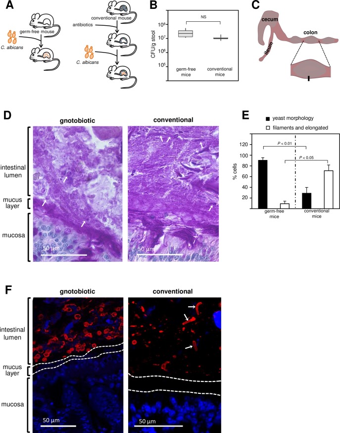
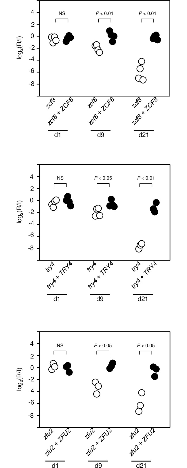
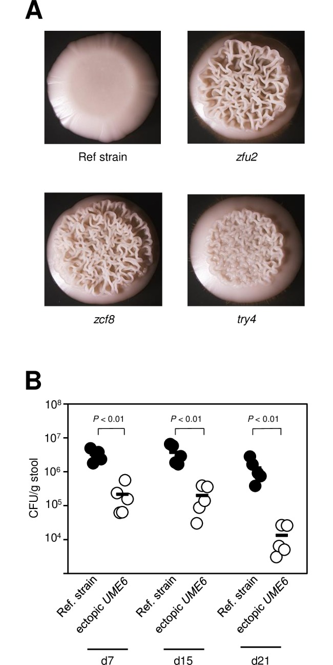
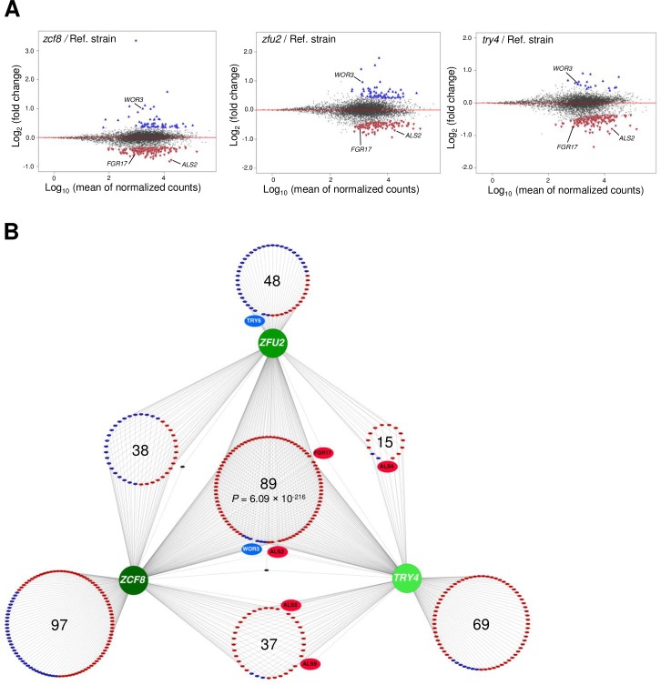
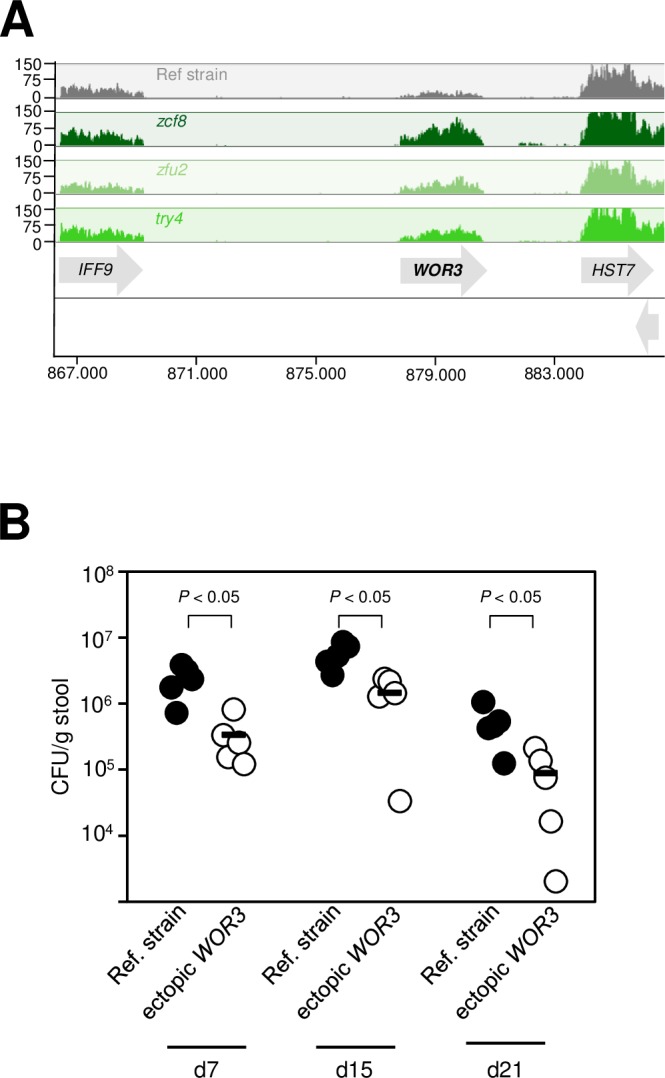
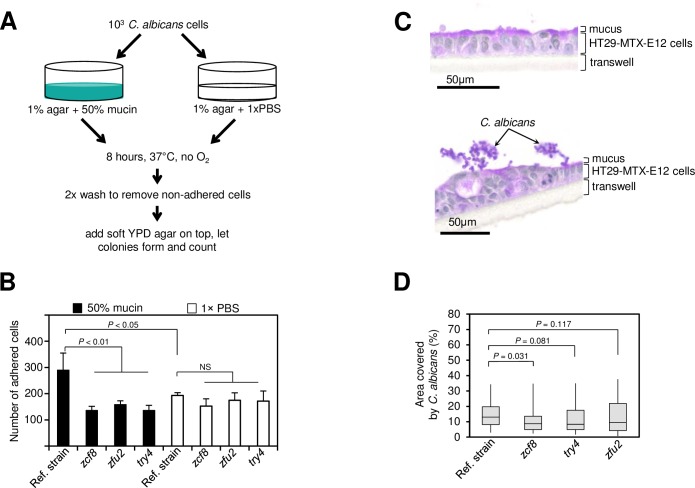
Many microorganisms that cause systemic, life-threatening infections in humans reside as harmless commensals in our digestive tract. Yet little is known about the biology of these microbes in the gut. Here, we visualize the interface between the human commensal and pathogenic fungus Candida albicans and the intestine of mice, a surrogate host. Because the indigenous mouse microbiota restricts C. albicans settlement, we compared the patterns of colonization in the gut of germ free and antibiotic-treated conventionally raised mice. In contrast to the heterogeneous morphologies found in the latter, we establish that in germ free animals the fungus almost uniformly adopts the yeast cell form, a proxy of its commensal state. By screening a collection of C. albicans transcription regulator deletion mutants in gnotobiotic mice, we identify several genes previously unknown to contribute to in vivo fitness. We investigate three of these regulators-ZCF8, ZFU2 and TRY4-and show that indeed they favor the yeast form over other morphologies. Consistent with this finding, we demonstrate that genetically inducing non-yeast cell morphologies is detrimental to the fitness of C. albicans in the gut. Furthermore, the identified regulators promote adherence of the fungus to a surface covered with mucin and to mucus-producing intestinal epithelial cells. In agreement with this result, histology sections indicate that C. albicans dwells in the murine gut in close proximity to the mucus layer. Thus, our findings reveal a set of regulators that endows C. albicans with the ability to endure in the intestine through multiple mechanisms.


Conflict of interest statement

The authors have declared that no competing interests exist.

Figures

Fig 1
Fig 1. Visualizing the interface between the fungus Candida albicans and the intestine of germ free or antibiotic-treated conventionally raised mice.
(A) Diagram illustrating two approaches to achieve murine gut colonization by C. albicans. The fungus can colonize the gastrointestinal tract of germ free animals (left) or conventionally raised mice that have been treated with antibiotics (right). (B) Fungal load in colony-forming units (CFU) per gram of stool in gnotobiotic and conventional animals 21 days after gavage. Six animals per group were evaluated. Shown is the median (horizontal line in the middle of the box) with interquartile range (grey box) and the distance to the minimum and maximum values (whisker). There are no statistical differences between the two groups. (C) Cartoon illustrating the portion of the intestine of mice colonized with C. albicans taken for histology and immunohistochemistry analyses. (D) Periodic acid-Schiff (PAS) stained tissue section (corresponding to the black rectangle in C) detailing the interface between the intestinal mucosa and the lumen. PAS stains polysaccharides, including those on the surface of C. albicans, in purple-magenta color. Notice the abundant oval shapes (arrows point to three of them) in the intestinal lumen of gnotobiotic animals, which correspond to C. albicans. Arrows in the right panel point to the diverse morphologies of the fungus observed in conventional rodents: Filaments (top), elongated cells (top right) and yeast (middle and mid-right). (E) Quantification of C. albicans cell morphologies observed in germ free and in conventional mice. Between 100–500 cells were scored in each one of 3–5 animals per group. The percentage of yeast cells as well as the percentage of filaments and elongated cells are significantly different between germ-free and conventional rodents. (F) Tissue sections after staining with DAPI (blue) and an anti-Candida antibody (red). Dotted lines represent the boundaries of the mucus layer. The arrows in the right panel point to elongated or filamenting C. albicans cells. Images are representative of multiple sections prepared from at least three mice. Statistical analyses by the Mann-Whitney test in (B) and (E). NS, not significant.
Fig 2
Fig 2. Identification of C. albicans transcription regulators that govern gut colonization in gnotobiotic mice.
(A) Schematic representation of the screening approach to identify genetic determinants of in vivo fitness in the fungus C. albicans. Each one of four pools consisting of 15–18 signature-tagged strains was gavaged in four germ-free mice. The abundance of each strain in the inoculum and after recovery from fecal pellets was determined by qPCR with oligos complementary to the signature tags. (B) Log2 (recovered/inoculum) values for each deletion mutant at one or 21 days post gavage (each column represents one mouse). Color intensity indicates reduction (blue) or accumulation (red). The order in which the mutants are displayed reflect hierarchical clustering. The eight mutants that cluster at the top display a consistent pattern of reduction in abundance in all mice at day 21. Four of these deletion mutants (orf19.4941, orf19.2315, orf19.921 and orf19.4722) had previously been identified in a screening conducted in the antibiotic-promoted gut colonization murine model. The deletion mutants try4Δ/Δ [orf19.5975], zfu2Δ/Δ [orf19.6781], zcf8Δ/Δ [orf19.1718], and orf19.5910Δ/Δ cluster together with the previous group but had not been identified before in any other mouse setting. (C) Validation of the gut fitness defect phenotype of three newly identified genes. Germ free animals were gavaged with 1:1 mixtures of the wild-type reference strain and try4Δ/Δ, zfu2Δ/Δ or zcf8Δ/Δ. The abundance of each strain in the inoculum (I) and after recovery from fecal pellets (R) at day 7, 15 and 21 was determined by qPCR (strains were barcoded). The deletion mutant strains were progressively depleted from the fecal pellets relative to the wild-type reference strain (P values are indicated on top of each comparison). Each circle represents the measurement from one mouse. Statistical analysis by the Mann-Whitney test. NS, not significant.
Fig 3
Fig 3. Adding back a wild-type copy of the respective genes to the deletion mutant strains restores in vivo fitness.
Germ free animals were gavaged with 1:1 mixtures of each deletion mutant and the corresponding gene add-back strain in three or four mice. The abundance of each strain in the inoculum (I) and after recovery from fecal pellets (R) was estimated by qPCR at day 1, 9 and 21 post gavage (strains were barcoded). In contrast to the deletion mutants which were depleted over time from the fecal pellets, the add-back strains maintained their relative abundance (P values are indicated on top of each comparison). Each circle represents the measurement from one mouse. Statistical analysis by two-tailed unpaired t-test assuming unequal variance. NS, not significant.
Fig 4
Fig 4. Filamentation is negatively associated with colonization of the gut of germ free mice.
(A) The deletion mutants zfu2, zcf8 and try4 display wrinkling—a proxy for filamentation—when grown under conditions that normally do not induce filamentation in C. albicans. The wild-type reference strain and the three deletion mutants were spotted on YPD agar and incubated at 30°C for 48h. (B) Fungal load in the feces of germ-free animals gavaged with a 1:1 mixture of wild-type reference strain and a mutant ectopically expressing the filament-inducing regulator UME6. Five animals per group were evaluated. Each circle represents the colony-forming units (CFU) per gram of stool from one mouse. Dashes represent the mean. Statistical analyses by the Mann-Whitney test. P values are indicated on top of each comparison.
Fig 5
Fig 5. RNA-seq mapping of the transcriptional regulatory network controlled by the identified regulators ZCF8, ZFU2 and TRY4.
(A) Plots show the distribution of all analyzed transcripts (∼6100 genes; each dot represents one transcript) when comparing the indicated deletion mutant to the wild-type reference strain. Plotted is the log2 fold change in expression (Y-axis) as a function of the mean of the normalized read counts (X-axis) for each transcript. Colored triangles correspond to transcripts whose expression is activated (i.e. positive regulation) (red) or repressed (i.e. negative regulation) (blue) by each regulator at Padj < 0.01. (B) Network view of the full set of genes whose expression is influenced by the identified C. albicans transcription regulators. The nodes of the network (in shades of green) are transcription regulators identified in our genetic screen. Large circles are composed of small colored ellipses. Each one of the latter represents an individual target gene. Positive (i.e. activation) and negative (i.e. repression) regulation is indicated by red and blue colors, respectively. The two black small ellipses depict the two genes that show discordant regulation (i.e. they are positively controlled by one regulator and negatively regulated by another). The shown network is completely based on RNA-seq data generated in this study.
Fig 6
Fig 6. Ectopic expression of WOR3 reduces C. albicans fitness in the gut of gnotobiotic mice.
(A) WOR3, a gene that promotes opaque cell formation, is one of the top targets of regulation of ZCF8, ZFU2 and TRY4. Shown are segments of RNA-seq tracks on chromosome R corresponding to the wild-type reference strain (grey) and the indicated mutants (green). Notice that the higher WOR3 transcript levels in the deletion mutants indicates negative regulation (i.e. repression). (B) Fungal load in the feces of germ-free animals gavaged with a 1:1 mixture of wild-type reference strain and a mutant ectopically expressing the opaque-inducing regulator WOR3. Five animals per group were evaluated. Each circle represents the colony-forming units (CFU) per gram of stool from one mouse. Dashes represent the mean. Statistical analyses by the Mann-Whitney test. P values are indicated on top of each comparison.
Fig 7
Fig 7. The regulators ZCF8, ZFU2 and TRY4 promote adherence.
(A) Schematic representation of in vitro assay to probe the ability of C. albicans cells to attach to a surface covered with intestinal mucin. (B) Plotted is the number of C. albicans cells (wild-type reference strain or deletion mutants) that remain attached to a semisolid surface containing either mucin (black bars) or PBS (white bars). Bars represent the mean ± S.D of at least three independent experiments; statistical analysis by t-test as described in Materials and methods. (C) C. albicans cells assemble in clusters on top of mucus-producing human intestinal epithelial cells HT29-MTX-E12. Shown are PAS-stained sections of HT29-MTX-E12 cells after 21 days of growth on transwells. Uninfected cells (top) and cells incubated with C. albicans (bottom). (D) Quantification of the attachment of C. albicans (wild-type reference strain or the indicated deletion mutants) to HT29-MTX-E12 cells grown on transwells. Plotted is the percentage area on top of the monolayer that was covered by fungal cells (as described in Materials and methods). Shown is the median (horizontal line in the middle of the box) with interquartile range (grey box) and the distance to the minimum and maximum values (whisker). Statistical analysis by the Mann-Whitney test. P values are indicated on top of each comparison.

Similar articles

Cited by

References

    1. Chudnovskiy A, Mortha A, Kana V, Kennard A, Ramirez JD, et al. (2016) Host-Protozoan Interactions Protect from Mucosal Infections through Activation of the Inflammasome. Cell 167: 444–456 e414. doi: 10.1016/j.cell.2016.08.076 - DOI - PMC - PubMed
    1. Lukes J, Stensvold CR, Jirku-Pomajbikova K, Wegener Parfrey L (2015) Are Human Intestinal Eukaryotes Beneficial or Commensals? PLoS Pathog 11: e1005039 doi: 10.1371/journal.ppat.1005039 - DOI - PMC - PubMed
    1. Underhill DM, Iliev ID (2014) The mycobiota: interactions between commensal fungi and the host immune system. Nat Rev Immunol 14: 405–416. doi: 10.1038/nri3684 - DOI - PMC - PubMed
    1. Wheeler ML, Limon JJ, Bar AS, Leal CA, Gargus M, et al. (2016) Immunological Consequences of Intestinal Fungal Dysbiosis. Cell Host Microbe 19: 865–873. doi: 10.1016/j.chom.2016.05.003 - DOI - PMC - PubMed
    1. Kumamoto CA (2016) The Fungal Mycobiota: Small Numbers, Large Impacts. Cell Host Microbe 19: 750–751. doi: 10.1016/j.chom.2016.05.018 - DOI - PubMed