. 2017 Oct 19;50(12):e6733.
doi: 10.1590/1414-431X20176733.

Myostatin promotes distinct responses on protein metabolism of skeletal and cardiac muscle fibers of rodents

Affiliations

Myostatin promotes distinct responses on protein metabolism of skeletal and cardiac muscle fibers of rodents

L H Manfredi et al. Braz J Med Biol Res. .

Abstract

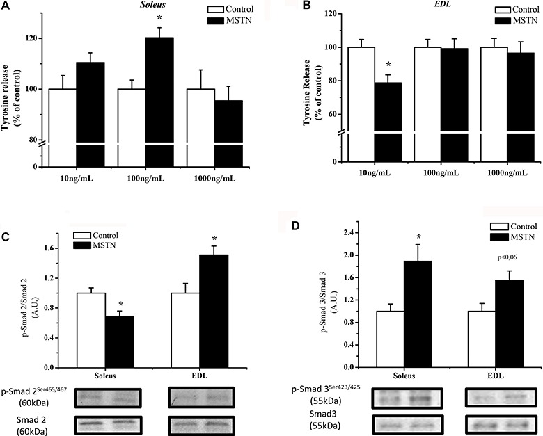
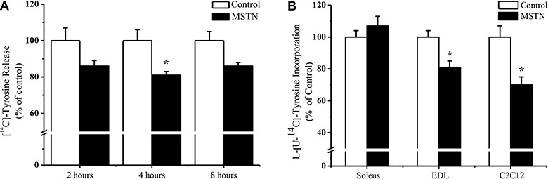
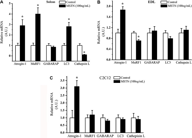
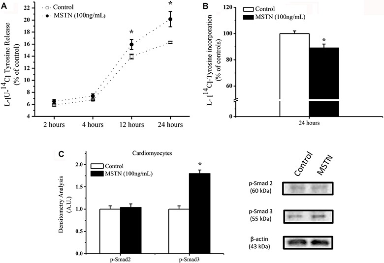
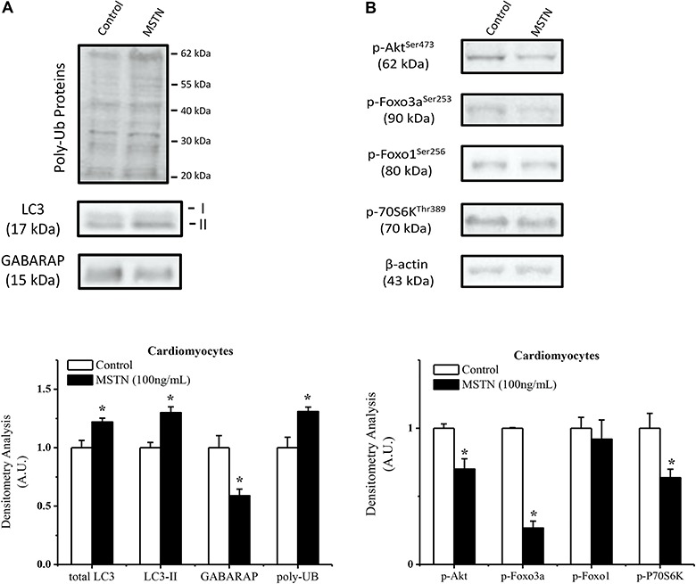
Myostatin is a novel negative regulator of skeletal muscle mass. Myostatin expression is also found in heart in a much less extent, but it can be upregulated in pathological conditions, such as heart failure. Myostatin may be involved in inhibiting protein synthesis and/or increasing protein degradation in skeletal and cardiac muscles. Herein, we used cell cultures and isolated muscles from rats to determine protein degradation and synthesis. Muscles incubated with myostatin exhibited an increase in proteolysis with an increase of Atrogin-1, MuRF1 and LC3 genes. Extensor digitorum longus muscles and C2C12 myotubes exhibited a reduction in protein turnover. Cardiomyocytes showed an increase in proteolysis by activating autophagy and the ubiquitin proteasome system, and a decrease in protein synthesis by decreasing P70S6K. The effect of myostatin on protein metabolism is related to fiber type composition, which may be associated to the extent of atrophy mediated effect of myostatin on muscle.


Figures

Figure 1.
Figure 1.. Overall proteolysis in (A) soleus and (B) extensor digitorum longus (EDL) muscles from rats incubated with myostatin (MSTN) for 4 h at different concentrations. Data are reported as means±SE (n=7). *P<0.05 MSTN vs Control, which is considered 100% (t-test). The control values of released tyrosine are (nmol·mg−1·2h−1): 0.302±0.016, 0.514±0.020, and 0.465±0.035 for soleus, and 0.268±0.012, 0.439±0.021, 0.370±0.020 for EDL at doses of 10, 100, and 1000 ng/mL, respectively. Phosphorylation status of Smad2 (C) and Smad3 (D) from soleus (100 ng/mL) and EDL (10 ng/mL) incubated with MSTN. A.U.: arbitrary units. *P<0.05 MSTN vs Control (t-test).
Figure 2.
Figure 2.. A, Overall proteolysis in C2C12 cells incubated with myostatin (MSTN) (100 ng/mL) after 2, 4, and 8 h. Data are reported as means±SE of labeled tyrosine released into the medium related to total amount of labeled tyrosine incorporated into proteins (%). Control was considered to be 100%, n=5. *P<0.05 MSTN vs Control (DMEM). B, Rate of protein synthesis in soleus and EDL muscles, and C2C12 cells incubated with MSTN at 100, 10, and 100 ng/mL, respectively, for 4 h. Data are reported as the means±SE of percentage of tyrosine incorporated into total protein (n=7). Control was considered to be 100%. *P<0.05 MSTN vs Control, t-test.
Figure 3.
Figure 3.. Gene expression of atrogin-1, MuRF1, GABARAP, LC3 and cathepsin L in A, soleus, B, extensor digitorum longus (EDL), and C, C2C12 cells incubated with myostatin (MSTN) for 4 h. Data are reported as the means±SE of percentages (n=6). Controls were considered to be 100%. *P<0.05 MSTN vs Control (t-test). A.U.: arbitrary units.
Figure 4.
Figure 4.. A, Overall proteolysis over time in cardiomyocytes incubated with myostatin (MSTN, 100 ng/mL). Data are reported as means±SE of the percentage of labeled tyrosine related to the total amount of labeled tyrosine incorporated into proteins. Control was considered to be 100%, n=6. B, Rate of protein synthesis in cardiomyocytes after 24 h of incubation with myostatin (100 ng/mL). Data are reported as means±SE of the percentage of tyrosine incorporated into total protein (n=7). Control was considered to be 100%. C, Phosphorylation status and densitometry analysis of Smad2 and Smad3 in cardiomyocytes incubated with myostatin for 4 h. Densitometry was corrected to β-actin. *P<0.05 MSTN vs Control (t-test).
Figure 5.
Figure 5.. A, Protein content of ubiquitinated (UB) proteins, total LC3 and form II, and GABARAP from cardiomyocytes incubated with myostatin (MSTN) (100 ng/mL) for 24 h with densitometry analysis of these proteins corrected to β-actin. Control was considered to be 1. Data are reported as means±SEM (n=5). B, Phosphorylation status of Akt, FoxO3a, FoxO1, P70S6K in cardiomyocytes incubated with myostatin for 24 h with densitometry analysis of these proteins corrected to their respective total proteins. Control was considered to be 1. Data are reported as means±SE (n=5). *P<0.05, t-test.

References

    1. McPherron AC, Lawler AM, Lee SJ. Regulation of skeletal muscle mass in mice by a new TGF-beta superfamily member. Nature. 1997;387:83–90. doi: 10.1038/387083a0. - DOI - PubMed
    1. Sharma M, Kambadur R, Matthews KG, Somers WG, Devlin GP, Conaglen JV, et al. Myostatin, a transforming growth factor-beta superfamily member, is expressed in heart muscle and is upregulated in cardiomyocytes after infarct. J Cell Physiol. 1999;180:1–9. doi: 10.1002/(SICI)1097-4652(199907)180:1<1::AID-JCP1>3.0.CO;2-V. - DOI - PubMed
    1. Trendelenburg AU, Meyer A, Rohner D, Boyle J, Hatakeyama S, Glass DJ. Myostatin reduces Akt/TORC1/p70S6K signaling, inhibiting myoblast differentiation and myotube size. Am J Physiol Cell Physiol. 2009;296:C1258–C1270. doi: 10.1152/ajpcell.00105.2009. - DOI - PubMed
    1. Zhang C, McFarlane C, Lokireddy S, Bonala S, Ge X, Masuda S, et al. Myostatin-deficient mice exhibit reduced insulin resistance through activating the AMP-activated protein kinase signalling pathway. Diabetologia. 2011;54:1491–1501. doi: 10.1007/s00125-011-2079-7. - DOI - PubMed
    1. Trendelenburg AU, Meyer A, Rohner D, Boyle J, Hatakeyama S, Glass DJ. Myostatin reduces Akt/TORC1/p70S6K signaling, inhibiting myoblast differentiation and myotube size. Am J Physiol Cell Physiol. 2009;296:C1258–C1270. doi: 10.1152/ajpcell.00105.2009. - DOI - PubMed

Publication types

MeSH terms