. 2017 May 11;8(42):71618-71629.
doi: 10.18632/oncotarget.17794. eCollection 2017 Sep 22.

CDK1 interacts with iASPP to regulate colorectal cancer cell proliferation through p53 pathway

Affiliations

CDK1 interacts with iASPP to regulate colorectal cancer cell proliferation through p53 pathway

Wei Gan et al. Oncotarget. .

Abstract

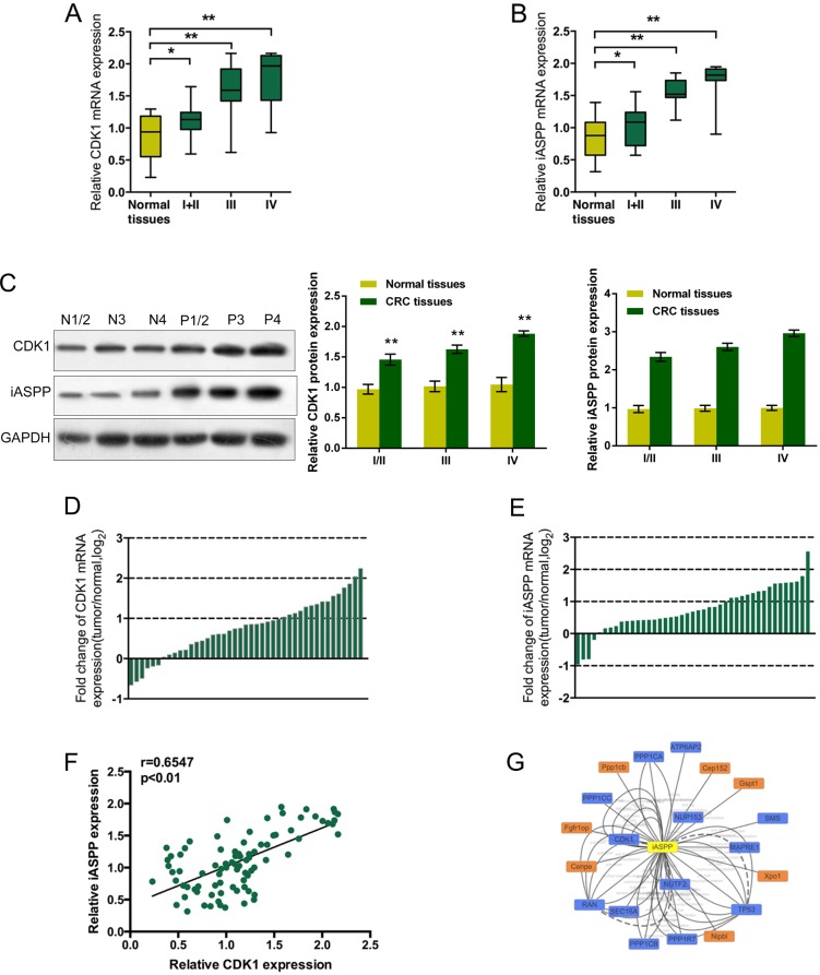
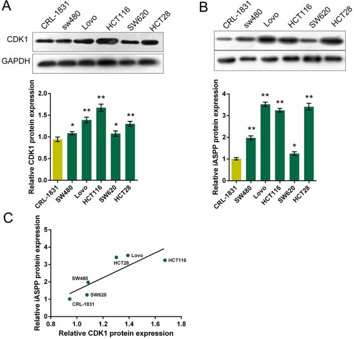
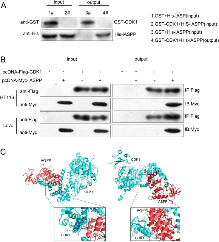
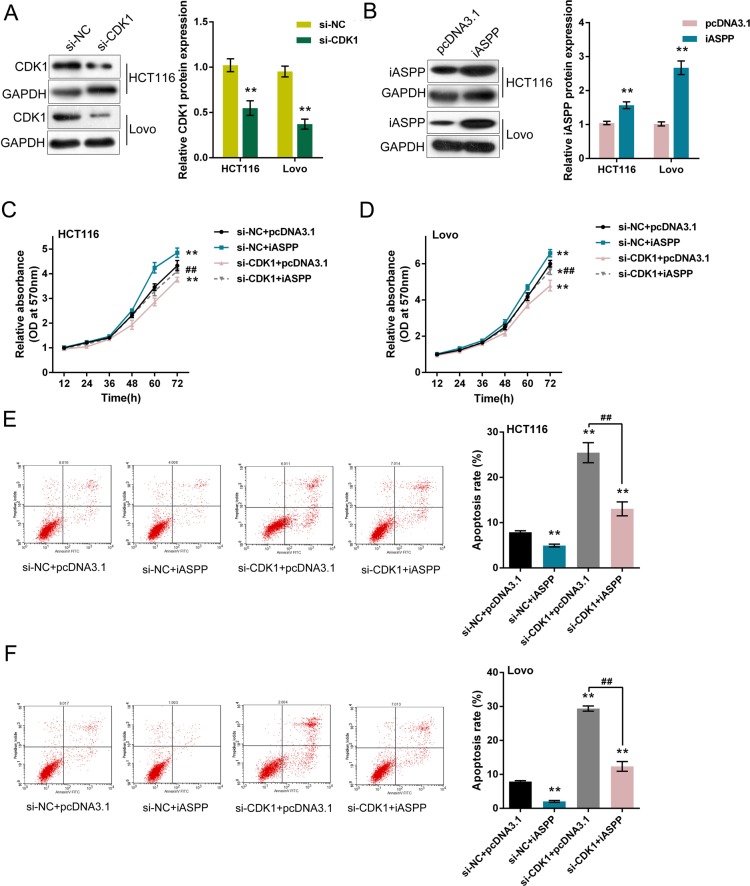
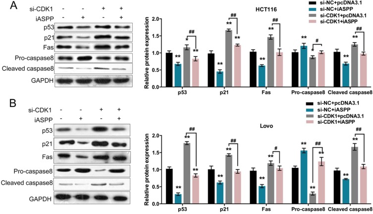
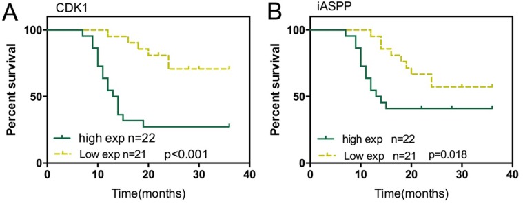
CDK1 (cyclin-dependent kinase 1) is a critical regulator of the G2-M checkpoint. CDK1 is considered a possible target for cancer treatment. In addition to CDK1, iASPP plays essential role in maintaining cancer cell proliferation. In the present study, we monitored the expression of CDK1 and iASPP at mRNA and protein levels in CRC tissues and cell lines; we also predicted that iASPP protein might interact with CDK1 protein. By performing GST pull-down assay and Co-IP assay, we confirmed the interaction of CDK1 and iASPP protein. In CRC cell lines, CDK1 interacted with iASPP to affect CRC cell proliferation and apoptosis; moreover, the p53 apoptosis pathway was involved in this progression. Taken together, we revealed that CDK1 and iASPP was up-regulated in CRC tissues and cell lines; CDK1 protein interacted with iASPP protein to affect CRC cell proliferation and apoptosis through the p53 apoptosis pathway. CDK1 and iASPP might serve as not only promising targets in CRC treatment, but also efficient prognostic markers. From the perspective of protein interactions, we provided a novel theoretical basis for targeted therapy of CRC.

Keywords: CDK1; cell proliferation; colorectal cancer; iASPP; p53.


Conflict of interest statement

CONFLICTS OF INTEREST None for all authors.

Figures

Figure 1
Figure 1
Expression levels of CDK1 and iASPP and their correlation in CRC tissues (A) and (B) The expression levels of CDK1 and iASPP mRNA in CRC tissues derived from patients in different stages were deteremined by using real-time PCR assays, compared to normal tissues. The data are presented as mean ± SD of three independent experiments. *P < 0.05, **P < 0.01. (C) The protein levels of CDK1 and iASPP in tumor tissues and adjacent normal tissues (normal tissues N1/2, N3 and N4; tumor tissues P1/2, P3 and P4) were determined using Western blot assays. (D) and (E) Expression of CDK1 and iASPP mRNA in 43 pairs of CRC tissues and their corresponding adjacent non-tumorous tissues (ANTs) in a training cohort. Expression level of CDK1 and iASPP mRNA was determined by real-time PCR and normalized to U6. Fold change were analyzed using the formula 2−(ΔΔCT [CRC/ ANT]). Red line indicates fold change of CDK1 or iASPP mRNA equal to 2. (F) The Spearman's rank correlation analysis was performed to analyze the correlation between CDK1 and iASPP mRNA in CRC tissues. (G) Data from Cytoscape showing the candidate proteins which could interact with iASPP protein.
Figure 2
Figure 2
Protein levels of CDK1 and iASPP in CRC cell lines (A) and (B) The protein levels of CDK1 and iASPP in five CRC cell lines, SW480, LoVo, HCT116, SW620 and HCT28 were determined using Western blot assays, compared to normal cell line, CRL-1831. The data are presented as mean ± SD of three independent experiments. *P < 0.05, **P < 0.01. (C) The Spearman's rank correlation analysis was performed to analyze the correlation between CDK1 and iASPP protein in CRC and normal cell lines.
Figure 3
Figure 3
Verification of the interaction between CDK1 and iASPP proteins (A) GST pull-down assays showing GST-CKD1 pulled down His-iASPP. (B) Confirmation of the interaction between CDK1 and iASPP using Co-IP assays. (C) The binding structure schematic diagram showing two possible interacting regions of CDK1 protein and iASPP protein, generated using PyMol.
Figure 4
Figure 4
Effect of co-processing CDK1 knockdown and iASPP overexpression on CRC cell proliferation and apoptosis (A) si-CDK1 was transfected into HCT116 and LoVo cells to achieve CDK1 knockdown, as verified by Western blot assays. (B) pcDNA3.1/iASPP was transfected into HCT116 and LoVo cells to achieve forced iASPP expression, as verified by Western blot assays. (C) and (D) si-CDK1 and pcDNA3.1/iASPP were co-transfected into HCT116 and LoVo cells; the cell viability was determined using MTT assays. (E) and (F) si-CDK1 and pcDNA3.1/iASPP were co-transfected into HCT116 and LoVo cells; the cell apoptosis was determined using Flow cytometer assays. The data are presented as mean ± SD of three independent experiments. *P < 0.05, **P < 0.01 (compared to control group), ##P < 0.01 (compared to si-CDK1 + pcDNA3.1 group.).
Figure 5
Figure 5
Reqirement of p53 pathway in CRC cell proliferation and apoptosis regulation (A) and (B) si-CDK1 and pcDNA3.1/iASPP were co-transfected into HCT116 and LoVo cells; the protein levels of p53, p21, Fas, Pro-caspase 8 and Cleaved caspase 8 were determined using Western blot assays. The data are presented as mean ± SD of three independent experiments. *P < 0.05, **P < 0.01 (compared to control group), ##P < 0.01 (compared to si-CDK1 + pcDNA3.1 group.).
Figure 6
Figure 6. Effect of iASPP and CDK1 expression on prognosis of CRC (A) Kaplan-Meier overall survival curves for 43 patients with CRC classified according to relative CDK1 expression level
(B) Kaplan-Meier overall survival curves for 43 patients with CRC classified according to relative iASPP expression level.

References

    1. Siegel R, Naishadham D, Jemal A. Cancer statistics, 2013. CA Cancer J Clin. 2013;63:11–30. - PubMed
    1. Wolpin BM, Meyerhardt JA, Mamon HJ, Mayer RJ. Adjuvant treatment of colorectal cancer. CA Cancer J Clin. 2007;57:168–185. - PubMed
    1. Huang WW, Ko SW, Tsai HY, Chung JG, Chiang JH, Chen KT, Chen YC, Chen HY, Chen YF, Yang JS. Cantharidin induces G2/M phase arrest and apoptosis in human colorectal cancer colo 205 cells through inhibition of CDK1 activity and caspase-dependent signaling pathways. Int J Oncol. 2011;38:1067–1073. - PubMed
    1. Sung WW, Lin YM, Wu PR, Yen HH, Lai HW, Su TC, Huang RH, Wen CK, Chen CY, Chen CJ, Yeh KT. High nuclear/cytoplasmic ratio of Cdk1 expression predicts poor prognosis in colorectal cancer patients. BMC Cancer. 2014;14:951. - PMC - PubMed
    1. Shapiro GI. Cyclin-dependent kinase pathways as targets for cancer treatment. J Clin Oncol. 2006;24:1770–1783. - PubMed