

Loss of Barx1 promotes hepatocellular carcinoma metastasis through up-regulating MGAT5 and MMP9 expression and indicates poor prognosis
- PMID: 29069753
- PMCID: PMC5641096
- DOI: 10.18632/oncotarget.18288
Loss of Barx1 promotes hepatocellular carcinoma metastasis through up-regulating MGAT5 and MMP9 expression and indicates poor prognosis
Abstract
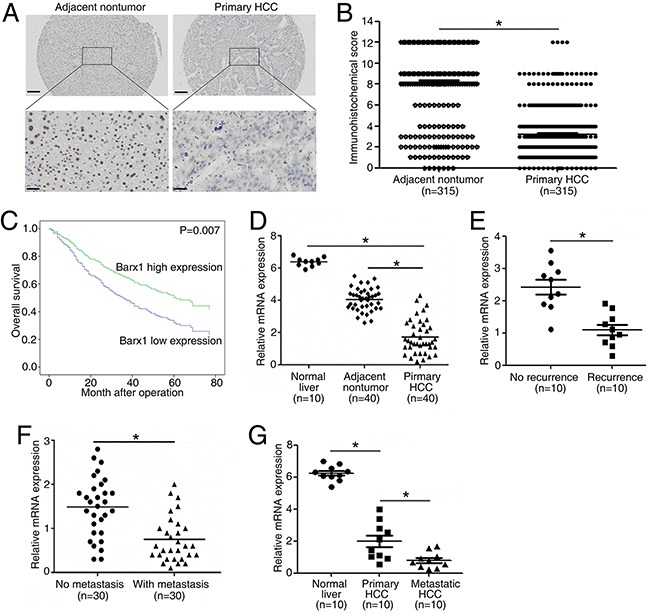
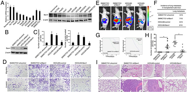
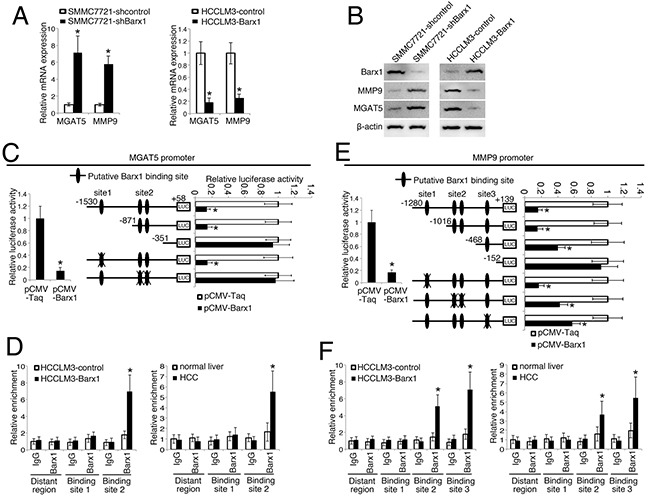
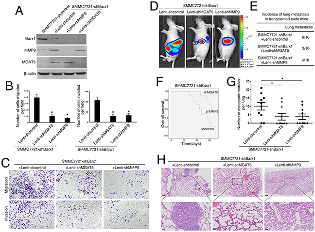
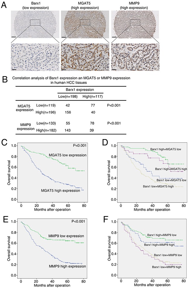
Metastasis is the major dominant reason for poor prognosis of hepatocellular carcinoma (HCC) after surgical treatment. However, the molecular mechanism of metastasis has not been well characterzied. Here, we report a novel function of Barx homeobox1 (Barx1) in inhibiting HCC invasion and metastasis. Barx1 expression is significantly decreased in human HCC tissues than in adjacent non-tumorous tissues and normal liver tissues. Low Barx1 expression is correlated with higher tumor-nodule-metastasis stage and indicates poor prognosis. Down-regulation of Barx1 promotes HCC migration, invasion and metastasis, whereas up-regulation of Barx1 inhibits HCC migration, invasion and metastasis. Mannosyl (alpha-1,6-)-glycoprotein beta-1,6-N-acetyl-glucosaminyltransferase 5 (MGAT5) and matrix metallopeptidase 9 (MMP9) are direct target genes of Barx1. Knockdown of Barx1 up-regulates MGAT5 and MMP9 expression in HCC cells with low metastatic capability, whereas over-expression of Barx1 suppresses their expression in HCC cells with high metastatic capability. Knockdown of both MGAT5 and MMP9 significantly decreases the invasion and metastasis abilities induced by Barx1 knockdown. Barx1 expression is negatively correlated with MGAT5 and MMP9 expression in human HCC tissues. Patients with low expression of Barx1 and high expression of MGAT5 or MMP9 are associated with poorer prognosis. Thus, loss of Barx1 represents a prognostic biomarker in human HCC patients.
Keywords: barx homeobox 1; hepatocellular carcinoma; mannosyl (alpha-1,6-)-glycoprotein beta-1,6-N-acetyl-glucosaminyltransferase 5; matrix metallopeptidase 9; metastasis.
Conflict of interest statement
CONFLICTS OF INTEREST The authors have no conflicts to disclose.
Figures
Similar articles
-
RUNX2 promotes hepatocellular carcinoma cell migration and invasion by upregulating MMP9 expression.Oncol Rep. 2016 Nov;36(5):2777-2784. doi: 10.3892/or.2016.5101. Epub 2016 Sep 19. Oncol Rep. 2016. PMID: 27666365
-
Fibulin-5 inhibits hepatocellular carcinoma cell migration and invasion by down-regulating matrix metalloproteinase-7 expression.BMC Cancer. 2014 Dec 12;14:938. doi: 10.1186/1471-2407-14-938. BMC Cancer. 2014. PMID: 25494879 Free PMC article.
-
IFITM3 promotes hepatocellular carcinoma invasion and metastasis by regulating MMP9 through p38/MAPK signaling.FEBS Open Bio. 2018 Jun 28;8(8):1299-1311. doi: 10.1002/2211-5463.12479. eCollection 2018 Aug. FEBS Open Bio. 2018. Retraction in: FEBS Open Bio. 2024 Mar;14(3):527. doi: 10.1002/2211-5463.13751. PMID: 30087833 Free PMC article. Retracted.
-
Overexpression of forkhead box C1 promotes tumor metastasis and indicates poor prognosis in hepatocellular carcinoma.Hepatology. 2013 Feb;57(2):610-24. doi: 10.1002/hep.26029. Hepatology. 2013. PMID: 22911555
-
Reduced expression of transcriptional intermediary factor 1 gamma promotes metastasis and indicates poor prognosis of hepatocellular carcinoma.Hepatology. 2014 Nov;60(5):1620-36. doi: 10.1002/hep.27273. Epub 2014 Sep 25. Hepatology. 2014. PMID: 24954480
Cited by
-
Aberrant N-glycosylation in cancer: MGAT5 and β1,6-GlcNAc branched N-glycans as critical regulators of tumor development and progression.Cell Oncol (Dordr). 2023 Jun;46(3):481-501. doi: 10.1007/s13402-023-00770-4. Epub 2023 Jan 23. Cell Oncol (Dordr). 2023. PMID: 36689079 Review.
-
Lipoprotein (a): a promising prognostic biomarker in patients with hepatocellular carcinoma after curative resection.Onco Targets Ther. 2018 Sep 17;11:5917-5924. doi: 10.2147/OTT.S164273. eCollection 2018. Onco Targets Ther. 2018. PMID: 30271176 Free PMC article.
-
Decreased miR-124-3p promoted breast cancer proliferation and metastasis by targeting MGAT5.Am J Cancer Res. 2019 Mar 1;9(3):585-596. eCollection 2019. Am J Cancer Res. 2019. PMID: 30949412 Free PMC article.
-
HAND1 and BARX1 Act as Transcriptional and Anatomic Determinants of Malignancy in Gastrointestinal Stromal Tumor.Clin Cancer Res. 2021 Mar 15;27(6):1706-1719. doi: 10.1158/1078-0432.CCR-20-3538. Epub 2021 Jan 15. Clin Cancer Res. 2021. PMID: 33451979 Free PMC article.
-
ZFP36 loss-mediated BARX1 stabilization promotes malignant phenotypes by transactivating master oncogenes in NSCLC.Cell Death Dis. 2023 Aug 16;14(8):527. doi: 10.1038/s41419-023-06044-z. Cell Death Dis. 2023. PMID: 37587140 Free PMC article.
References
-
- Forner A, Llovet JM, Bruix J. Hepatocellular carcinoma. The Lancet. 2012;379:1245–1255. - PubMed
-
- El-Serag HB. Hepatocellular carcinoma. The New England journal of medicine. 2011;365:1118–1127. - PubMed
-
- Gould DB, Walter MA. Cloning, characterization, localization, and mutational screening of the human BARX1 gene. Genomics. 2000;68:336–342. - PubMed
-
- Tissier-Seta JP, Mucchielli ML, Mark M, Mattei MG, Goridis C, Brunet JF. Barx1, a new mouse homeodomain transcription factor expressed in cranio-facial ectomesenchyme and the stomach. Mechanisms of development. 1995;51:3–15. - PubMed
-
- Kim BM, Buchner G, Miletich I, Sharpe PT, Shivdasani RA. The stomach mesenchymal transcription factor Barx1 specifies gastric epithelial identity through inhibition of transient Wnt signaling. Developmental cell. 2005;8:611–622. - PubMed
LinkOut - more resources
Full Text Sources
Other Literature Sources
Research Materials
Miscellaneous
