. 2017 Aug 7;8(42):72167-72181.
doi: 10.18632/oncotarget.20050. eCollection 2017 Sep 22.

Small-molecule inhibitors of PD-1/PD-L1 immune checkpoint alleviate the PD-L1-induced exhaustion of T-cells

Affiliations

Small-molecule inhibitors of PD-1/PD-L1 immune checkpoint alleviate the PD-L1-induced exhaustion of T-cells

Lukasz Skalniak et al. Oncotarget. .

Abstract

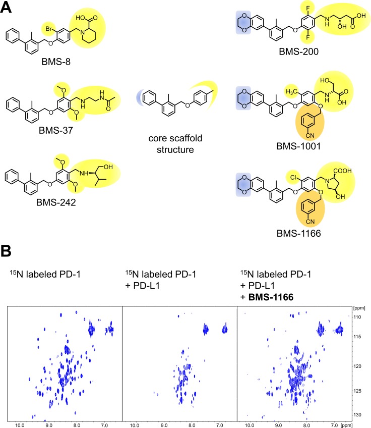
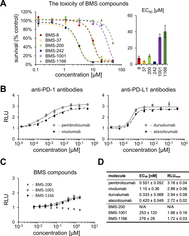
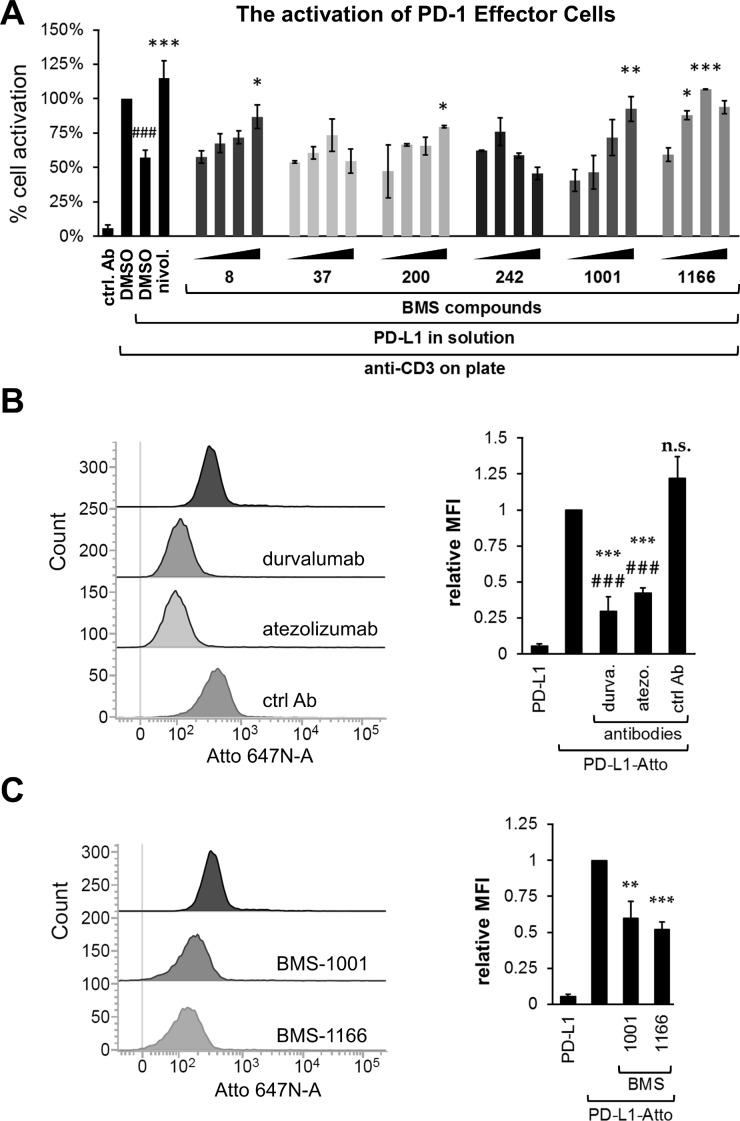
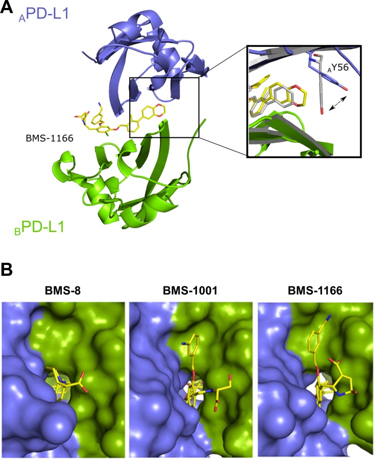
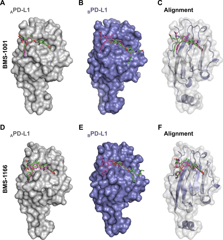
Antibodies targeting the PD-1/PD-L1 immune checkpoint achieved spectacular success in anticancer therapy in the recent years. In contrast, no small molecules with cellular activity have been reported so far. Here we provide evidence that small molecules are capable of alleviating the PD-1/PD-L1 immune checkpoint-mediated exhaustion of Jurkat T-lymphocytes. The two optimized small-molecule inhibitors of the PD-1/PD-L1 interaction, BMS-1001 and BMS-1166, developed by Bristol-Myers Squibb, bind to human PD-L1 and block its interaction with PD-1, when tested on isolated proteins. The compounds present low toxicity towards tested cell lines and block the interaction of soluble PD-L1 with the cell surface-expressed PD-1. As a result, BMS-1001 and BMS-1166 alleviate the inhibitory effect of the soluble PD-L1 on the T-cell receptor-mediated activation of T-lymphocytes. Moreover, the compounds were effective in attenuating the inhibitory effect of the cell surface-associated PD-L1. We also determined the X-ray structures of the complexes of BMS-1001 and BMS-1166 with PD-L1, which revealed features that may be responsible for increased potency of the compounds compared to their predecessors. Further development may lead to the design of an anticancer therapy based on the orally delivered immune checkpoint inhibition.

Keywords: PD-1; PD-L1; immune checkpoint blockade; inhibitor; small-molecules.


Conflict of interest statement

CONFLICTS OF INTEREST The authors declare no conflicts of interests.

Figures

Figure 1
Figure 1. Structures and the PD-1/PD-L1 blocking potential of BMS compounds
(A) The structures of BMS compounds tested in this study. The original numbering found in the patents [24, 25] is used. (B) BMS-1166 dissociates a preformed PD-1/PD-L1 complex, as shown by the NMR-based Antagonist Induced Dissociation Assay (AIDA). 1H-15N HMQC spectra are shown for the 15N labeled PD-1 (left) and the complex of the 15N labeled PD-1 and unlabeled PD-L1 alone (center) and after addition of BMS-1166 (right). Linewidth broadening, observed as loss of resonance signals in the central panel, indicates increased transverse relaxation rates associated with the complex formation. At the PD-L1:BMS-1166 molar ratio of 4:1, BMS-1166 efficiently disrupted the PD-1/PD-L1 complex, as visualized by the restoration of the 1H-15N signals of PD-1.
Figure 2
Figure 2. Cytotoxicity and activity of BMS compounds in PD-1/PD-L1 checkpoint assay
(A) Cytotoxicity of BMS compounds against the PD-1 Effector Cells was tested using metabolic activity assay following the 48 h treatment of the cells with the indicated compounds. The presented EC50 values (right panel) are means ± SEM from the three independent experiments (left panel shows a representative result). (B, C) Activities of reference antibodies (B) and BMS compounds (C) in alleviating the effect of PD1/PD-L1 checkpoint on TCR-mediated T cell activation are expressed as the level of luciferase activity (for details see Materials and Methods). The graphs present relative luminescence normalized to the DMSO-treated controls and are representative of the three independent experiments. (D) The result of the fitting of Hill model to experimental data presented on panels B and C. EC50 values represent half maximal effective concentrations, and RLUmax values represent maximal relative luminescence values, and illustrate the potency of the listed molecules in restoring the activity of ECs in the assay.
Figure 3
Figure 3. BMS compounds restore the sPD-L1-supressed activation of Jurkat T-cells
(A) Effector Cells were activated for 24 h with the anti-CD3 antibody alone or in the presence of sPD-L1, pre-incubated with nivolumab (nivol.), DMSO or the BMS compounds. The activity of luciferase, expressed in response to TCR-mediated induction of NFAT-responsive promoter, was determined as an indicator of cell activation. BMS compounds dose-dependently restored the activation, pre-blocked by the presence of sPD-L1. The graphs present mean ± SEM from at least three independent experiments. Statistical significance was evaluated using a one-way ANOVA with the Tukey's post-hoc test: *p < 0.05, **p < 0.01, ***p < 0.001. (B, C) The binding of the fluorescently-labeled sPD-L1 (PD-L1-Atto) to PD-1 expressing cells determined using flow cytometry. PD-L1-Atto was pre-incubated with tested compounds prior to staining. Cell staining is blocked in the presence of the anti-PD-L1 antibody (durvalumab, durva. or atezolizumab, atezo.) and BMS compounds. MFI – Geo Mean Fluorescence Intensity values. The bar graphs present mean ± SEM from three independent experiments. For the statistics, t-test was used: **p < 0.01, ***p < 0.001.
Figure 4
Figure 4. BMS-1166 induces binding cleft opening
(A) Arrangement of the molecules in the crystal structure – two PD-L1 molecules form a single pocket accommodating BMS compound. A close-up presents the position of the side-chain of the Tyr56 in BMS-8 (gray) and BMS-1166 (yellow/blue) containing structures. (B) The 2,3-dihydro-1,4-benzodioxine moiety of BMS-1001 and BMS-1166 induces the previously absent side-chain movement that triggers transformation of the binding pocket into the binding tunnel across the transverse vertical axis of the dimer. The panel visualizes surface representation of the dimer with the visible binding cleft (left, BMS-8/PD-L1 complex) and the binding tunnel (center and right for the BMS-1001/PD-L1 and BMS-1166/PD-L1 complexes, respectively). PD-L1 molecules forming the dimer are colored blue and green for chains A and B, respectively. Compounds are shown as yellow sticks.
Figure 5
Figure 5. Decomposition of BMS-1166
BMS-1166 (1) or its fragments (2-6) were tested for the interaction with PD-L1 (blue) using 1H-15N HMQC NMR method. The spectra determined in presence of compounds (1)-(4) display line-broadening (red), indicative of compound-induced formation of a higher molecular weight PD-L1 complex. Compounds (5) and (6) induced no changes in the spectra even at molar excess, indicating the lack of interaction.
Figure 6
Figure 6. The prediction of BMS-1001 and −1166 binding sites on PD-L1 surface
Molecular docking simulations were performed to predict the favored binding sites of BMS compounds to PD-L1 protein. APD-L1 and BPD-L1 protomers were extracted from the co-crystal structures of BMS-1001/PD-L1 and BMS-1166/PD-L1 and used as templates (receptors) for the docking of appropriate BMS compound (ligand). The best docking results (represented by the purple compounds) are compared with crystallographic arrangements (represented by the green compounds). (A, D) For both BMS compounds, when APD-L1 protomer was investigated, the preferred molecular docking results corresponded well to the binding modes determined from crystallographic data. (B, E) When BPD-L1 protomer was selected, molecular docking simulations suggested an opposite orientation of the compounds to the one defined by crystal structure analysis (among the 10 best results not a single resembled the BMS orientation determined from crystal structures of BMS/BPD-L1). (C, F) The alignment of BPD-L1 protomer to the APD-L1 protomer revealed a significant resemblance of the binding mode of BMS compounds docked to BPD-L1 and the ones revealed from the crystals for APD-L1 protomer.

References

    1. Sharma P, Allison JP. Immune checkpoint targeting in cancer therapy: toward combination strategies with curative potential. Cell. 2015;161:205–14. doi: 10.1016/j.cell.2015.03.030. - DOI - PMC - PubMed
    1. Hoos A. Development of immuno-oncology drugs–from CTLA4 to PD1 to the next generations. Nat Rev Drug Discov. 2016;15:235–47. doi: 10.1038/nrd.2015.35. - DOI - PubMed
    1. Topalian SL, Drake CG, Pardoll DM. Immune checkpoint blockade: a common denominator approach to cancer therapy. Cancer Cell. 2015;27:450–61. doi: 10.1016/j.ccell.2015.03.001. - DOI - PMC - PubMed
    1. Mahoney KM, Rennert PD, Freeman GJ. Combination cancer immunotherapy and new immunomodulatory targets. Nat Rev Drug Discov. 2015;14:561–84. doi: 10.1038/nrd4591. - DOI - PubMed
    1. Khalil DN, Smith EL, Brentjens RJ, Wolchok JD. The future of cancer treatment: immunomodulation, CARs and combination immunotherapy. Nat Rev Clin Oncol. 2016;13:273–90. doi: 10.1038/nrclinonc.2016.25. - DOI - PMC - PubMed