

Hemorrhagic hypopyon as presenting feature of intravascular lymphoma, a case report
- PMID: 29070018
- PMCID: PMC5657083
- DOI: 10.1186/s12886-017-0591-3
Hemorrhagic hypopyon as presenting feature of intravascular lymphoma, a case report
Abstract
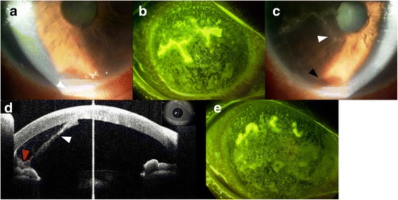
Background: Herpes uveitis has been previously reported to present with hyphema, but hemorrhagic hypopyon is rarely reported as a herpetic uveitis manifestation. We report a case of herpes simplex virus (HSV) presenting with hemorrhagic hypopyon, and speculate on the underlying pathophysiology with relation to an intravascular lymphoma which was subsequently diagnosed as a result.
Case presentation: We present a case wherein a 62-year-old Japanese rheumatoid arthritis woman, with HSV uveitis, presented with hemorrhagic hypopyon in the anterior chamber and a fever with photophobia. Patient was treated with antiviral drugs which improved the hyphema and corneal lesions, but lesions recurred 3 months later. This rare presentation of HSV induced uveitis, and its subsequent recurrence, aroused suspicion of an additional hypopyon-inducing pathology. On account of previous history of lung opacities and elevated LDH, intravascular lymphoma was eventually diagnosed via lung biopsy. She was treated for the lymphoma which also completely resolved all ocular symptoms without any recurrence as of 1.5 years later.
Conclusion: The exceedingly rare presentation of hemorrhagic hypopyon may have been enabled by an interaction of the HSV with the intravascular lymphoma. HSV involvement was indicated by the dendritic lesions, IgG assay, and response to anti-viral drugs. The ocular involvement of the intravascular lymphoma seems to be indicated by virtue of the anti-tumor drugs completely resolving all ocular symptoms.
Keywords: Case report; Hemorrhagic hypopyon; Herpes simplex uveitis; Intravascular lymphoma; Ophthalmic manifestations; Vessel permeability.
Conflict of interest statement
Ethics approval and consent to participate
The Institutional Review Board of the Osaka University Medical School approved the research protocol, and the procedures conformed to the tenets of the Declaration of Helsinki.
Consent for publication
Written informed consent was obtained from the patient for publication of this Case report and any accompanying images. A copy of the written consent is available for review by the Editor of this journal.
Competing interests
The authors declare that they have no competing interests.
Publisher’s Note
Springer Nature remains neutral with regard to jurisdictional claims in published maps and institutional affiliations.
Figures


References
-
- Biswas J, Samanta TK, Madhavan HN, Kumarasamy N, Solomon S. Acute panuveitis with haemorrhagic hypopyon as a presenting feature of acquired immunodeficiency syndrome (AIDS) Indian J Ophthalmol. 2000;48:311–312. - PubMed
-
- Verity DH, Graham EM, Carr R, van der Walt JD, Stanford MR. Hypopyon uveitis and iris nodules in non-Hodgkin's lymphoma: ocular relapse during systemic remission. Clin Oncol (R Coll Radiol) 2000;12:292–294. - PubMed
Publication types
MeSH terms
LinkOut - more resources
Full Text Sources
Other Literature Sources
Medical
