. 2017 Oct 25;14(1):208.
doi: 10.1186/s12974-017-0984-5.

Human antibodies against the myelin oligodendrocyte glycoprotein can cause complement-dependent demyelination

Affiliations

Human antibodies against the myelin oligodendrocyte glycoprotein can cause complement-dependent demyelination

Patrick Peschl et al. J Neuroinflammation. .

Abstract

Background: Antibodies to the myelin oligodendrocyte glycoprotein (MOG) are associated with a subset of inflammatory demyelinating diseases of the central nervous system such as acute disseminated encephalomyelitis and neuromyelitis optica spectrum disorders. However, whether human MOG antibodies are pathogenic or an epiphenomenon is still not completely clear. Although MOG is highly conserved within mammals, previous findings showed that not all human MOG antibodies bind to rodent MOG. We therefore hypothesized that human MOG antibody-mediated pathology in animal models may only be evident using species-specific MOG antibodies.

Methods: We screened 80 human MOG antibody-positive samples for their reactivity to mouse and rat MOG using either a live cell-based assay or immunohistochemistry on murine, rat, and human brain tissue. Selected samples reactive to either human MOG or rodent MOG were subsequently tested for their ability to induce complement-mediated damage in murine organotypic brain slices or enhance demyelination in an experimental autoimmune encephalitis (EAE) model in Lewis rats. The MOG monoclonal antibody 8-18-C5 was used as a positive control.

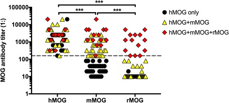
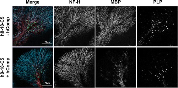
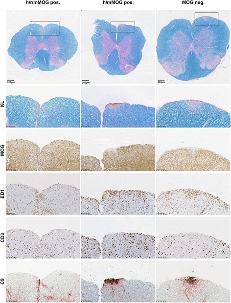
Results: Overall, we found that only a subset of human MOG antibodies are reactive to mouse (48/80, 60%) or rat (14/80, 18%) MOG. Purified serum antibodies from 10 human MOG antibody-positive patients (8/10 reactive to mouse MOG, 6/10 reactive to rat MOG), 3 human MOG-negative patients, and 3 healthy controls were tested on murine organotypic brain slices. Purified IgG from one patient with high titers of anti-human, mouse, and rat MOG antibodies and robust binding to myelin tissue produced significant, complement-mediated myelin loss in organotypic brain slices, but not in the EAE model. Monoclonal 8-18-C5 MOG antibody caused complement-mediated demyelination in both the organotypic brain slice model and in EAE.

Conclusion: This study shows that a subset of human MOG antibodies can induce complement-dependent pathogenic effects in a murine ex vivo animal model. Moreover, a high titer of species-specific MOG antibodies may be critical for demyelinating effects in mouse and rat animal models. Therefore, both the reactivity and titer of human MOG antibodies must be considered for future pathogenicity studies.

Keywords: Antibodies; EAE; MOG; Myelin oligodendrocyte glycoprotein; Neuromyelitis optica spectrum disorders; Organotypic slice culture.


Conflict of interest statement

Ethics approval and consent to participate

This study was approved by the Ethical Committee of the Medical University of Innsbruck (study numbers AM3041A and AM4059) and by the Ethic Committees of the Hospital Clinic of Barcelona (2010/5680), Charite University Medicine Berlin (EA1/131/09) and University Hospital Zürich (KEK-Nr. 2013-0001). All patients or their caregivers gave informed written consent.

Consent for publication

Not applicable.

Competing interests

The Medical University of Innsbruck and University Hospital Innsbruck and Medical University of Vienna receive payments for antibody assays (aquaporin-4 and other anti-neuronal and anti-glial antibodies) and for aquaporin-4 antibody validation assays organized by Euroimmun (Lübeck, Germany).

Publisher’s Note

Springer Nature remains neutral with regard to jurisdictional claims in published maps and institutional affiliations.

Figures

Fig. 1
Fig. 1
Serum antibody titer levels to human, mouse, and rat MOG by live cell-based assay. The cutoff titer value of 1:160 is indicated by a dashed horizontal line. Individual antibody titers are shown as black circles (reactive to hMOG only), yellow triangles (reactive to hMOG+mMOG) or red squares (reactive to hMOG+mMOG+rMOG). Antibody titers were compared using a non-parametric test (Friedman’s test with Dunn’s multiple comparisons test, overall p value < 0.001). ***p < 0.001
Fig. 2
Fig. 2
a Myelin staining with serum from human samples and monoclonal 8-18-C5 on human, mouse, or rat cerebellum. White matter tracts and folia were clearly stained with MOG antibody-positive serum samples and 8-18-C5 but not in MOG antibody-negative control samples. Sections were magnified at ×20 (mouse/rat tissue) and at ×10 (human tissue). b Serum samples reactive to human and mouse MOG bind to both human and mouse cerebellum sections. Pre-adsorbed samples show a significant reduction in MOG-specific myelin staining. Sections were magnified at ×2.5 (human tissue) and at ×20 (mouse tissue). Inlays show higher magnification (10×) of human white matter tracts
Fig. 3
Fig. 3
Murine organotypic brain slices incubated with human monoclonal 8-18-C5 with (+ hComp) and without (− hComp) human complement. Cerebellar folia of proteolipid protein (PLP)-EGFP transgenic mice are stained for neurofilament-heavy (NF-H, blue) and myelin basic protein (MBP; red). Confocal images were taken with ×25 objectives
Fig. 4
Fig. 4
One of 10 human MOG antibody samples causes mild demyelination in murine cerebellar slice culture. Murine organotypic brain slices incubated with total IgG purified from MOG-positive (MOG pos.) and MOG-negative (MOG neg.) human sera with human complement (+hComp) were stained for neurofilament-heavy (NF-H; blue), calbindin (blue), and myelin basic protein (MBP; red). Mild loss of MBP was observed only in the presence of MOG pos. serum (MOG 7) and hComp. Confocal images were taken with ×25 objectives and ×3 digital zoom for higher magnifications
Fig. 5
Fig. 5
Spinal cord sections of MBP-T cell EAE animals co-injected with patient antibodies. Patient IgG was reactive to either human, rat, and mouse MOG (h/r/mMOG pos., MOG 7); human and mouse MOG (h/mMOG pos., MOG 6); or negative to human MOG (hMOG neg.) tested by CBA KL, Kluver-Barrera (myelin); MOG, myelin oligodendrocyte glycoprotein; ED1, macrophages; CD3, T cells; C9, complement. Spinal cord slices were digitally magnified at 3.8× and highlighted sections at ×20
Fig. 6
Fig. 6
EAE animals co-injected with mouse monoclonal antibody 8-18-C5 (m8-18-C5) vs. isotype control (isotype Ctrl.). Slices were stained with KL, Kluver-Barrera (myelin); ED1, macrophages; C9, complement; CD3, T cells. Spinal cord slices were digitally magnified at ×3.8

References

    1. O'Connor KC, et al. Self-antigen tetramers discriminate between myelin autoantibodies to native or denatured protein. Nat Med. 2007;13(2):211–217. doi: 10.1038/nm1488. - DOI - PMC - PubMed
    1. Brilot F, et al. Antibodies to native myelin oligodendrocyte glycoprotein in children with inflammatory demyelinating central nervous system disease. Ann Neurol. 2009;66(6):833–842. doi: 10.1002/ana.21916. - DOI - PubMed
    1. Selter RC, et al. Antibody responses to EBV and native MOG in pediatric inflammatory demyelinating CNS diseases. Neurology. 2010;74(21):1711–1715. doi: 10.1212/WNL.0b013e3181e04096. - DOI - PubMed
    1. Probstel AK, et al. Antibodies to MOG are transient in childhood acute disseminated encephalomyelitis. Neurology. 2011;77(6):580–588. doi: 10.1212/WNL.0b013e318228c0b1. - DOI - PubMed
    1. Lalive PH, et al. Highly reactive anti-myelin oligodendrocyte glycoprotein antibodies differentiate demyelinating diseases from viral encephalitis in children. Mult Scler. 2011;17(3):297–302. doi: 10.1177/1352458510389220. - DOI - PubMed

MeSH terms

LinkOut - more resources