Adenosine stress CMR T1-mapping detects early microvascular dysfunction in patients with type 2 diabetes mellitus without obstructive coronary artery disease
- PMID: 29070069
- PMCID: PMC5655826
- DOI: 10.1186/s12968-017-0397-8
Adenosine stress CMR T1-mapping detects early microvascular dysfunction in patients with type 2 diabetes mellitus without obstructive coronary artery disease
Erratum in
-
Correction to: Adenosine stress CMR T1-mapping detects early microvascular dysfunction in patients with type 2 diabetes mellitus without obstructive coronary artery disease.J Cardiovasc Magn Reson. 2017 Dec 7;19(1):99. doi: 10.1186/s12968-017-0406-y. J Cardiovasc Magn Reson. 2017. PMID: 29212500 Free PMC article.
Abstract
Background: Type 2 diabetes mellitus (T2DM) is associated with coronary microvascular dysfunction in the absence of obstructive coronary artery disease (CAD). Cardiovascular magnetic resonance (CMR) T1-mapping at rest and during adenosine stress can assess coronary vascular reactivity. We hypothesised that the non-contrast T1 response to vasodilator stress will be altered in patients with T2DM without CAD compared to controls due to coronary microvascular dysfunction.
Methods: Thirty-one patients with T2DM and sixteen matched healthy controls underwent CMR (3 T) for cine, rest and adenosine stress non-contrast T1-mapping (ShMOLLI), first-pass perfusion and late gadolinium enhancement (LGE) imaging. Significant CAD (>50% coronary luminal stenosis) was excluded in all patients by coronary computed tomographic angiography.
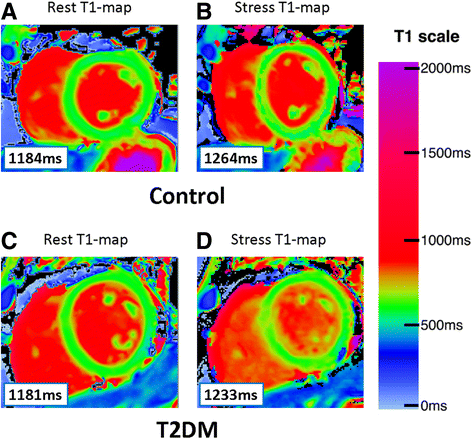
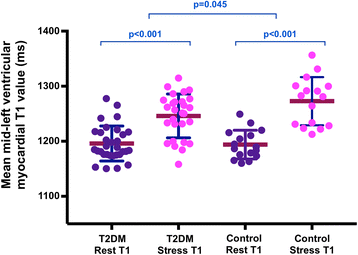
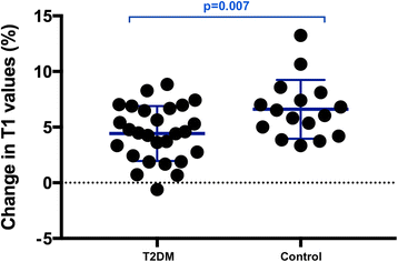
Results: All subjects had normal left ventricular (LV) ejection and LV mass index, with no LGE. Myocardial perfusion reserve index (MPRI) was lower in T2DM than in controls (1.60 ± 0.44 vs 2.01 ± 0.42; p = 0.008). There was no difference in rest native T1 values (p = 0.59). During adenosine stress, T1 values increased significantly in both T2DM patients (from 1196 ± 32 ms to 1244 ± 44 ms, p < 0.001) and controls (from 1194 ± 26 ms to 1273 ± 44 ms, p < 0.001). T2DM patients showed blunted relative stress non-contrast T1 response (T2DM: ΔT1 = 4.1 ± 2.9% vs.
Controls: ΔT1 = 6.6 ± 2.6%, p = 0.007) due to a blunted maximal T1 during adenosine stress (T2DM 1244 ± 44 ms vs. controls 1273 ± 44 ms, p = 0.045).
Conclusions: Patients with well controlled T2DM, even in the absence of arterial hypertension and significant CAD, exhibit blunted maximal non-contrast T1 response during adenosine vasodilatory stress, likely reflecting coronary microvascular dysfunction. Adenosine stress and rest T1 mapping can detect subclinical abnormalities of the coronary microvasculature, without the need for gadolinium contrast agents. CMR may identify early features of the diabetic heart phenotype and subclinical cardiac risk markers in patients with T2DM, providing an opportunity for early therapeutic intervention.
Keywords: Cardiovascular magnetic resonance; Diabetes mellitus; Microvascular obstruction; Myocardial perfusion; ShMOLLI T1-mapping.
Conflict of interest statement
Ethics approval and consent to participate
The study was approved by the National Research Ethics Committee (Ref 13/SW/0257), and informed written consent was obtained from each participant.
Consent for publication
All subjects gave written informed consent for publication.
Competing interests
SKP has patent authorship rights for U.S. patent 9,285,446 B2. Systems and methods for shortenedLlook Locker inversion recovery (Sh-MOLLI) cardiac gated mapping of T1. Granted March 15, 2016. All rights transferred to Siemens Medical.
All other authors have no relationships relevant to the contents of this paper to disclose.
Publisher’s Note
Springer Nature remains neutral with regard to jurisdictional claims in published maps and institutional affiliations.
Figures


References
Publication types
MeSH terms
Substances
Grants and funding
LinkOut - more resources
Full Text Sources
Other Literature Sources
Medical
Miscellaneous

