. 2017 Dec;175(4):1839-1852.
doi: 10.1104/pp.17.01152. Epub 2017 Oct 25.

Phosphorylation of Arabidopsis MAP Kinase Phosphatase 1 (MKP1) Is Required for PAMP Responses and Resistance against Bacteria

Affiliations

Phosphorylation of Arabidopsis MAP Kinase Phosphatase 1 (MKP1) Is Required for PAMP Responses and Resistance against Bacteria

Lingyan Jiang et al. Plant Physiol. 2017 Dec.

Abstract

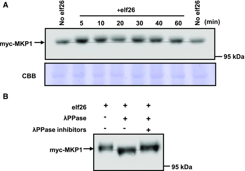
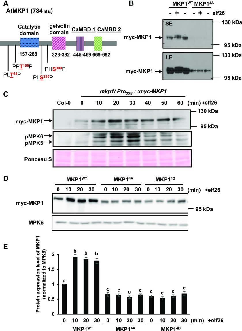
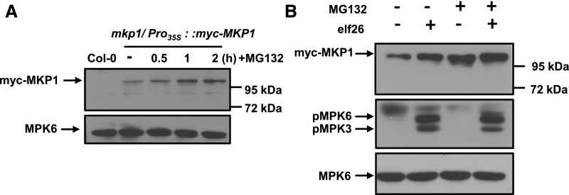
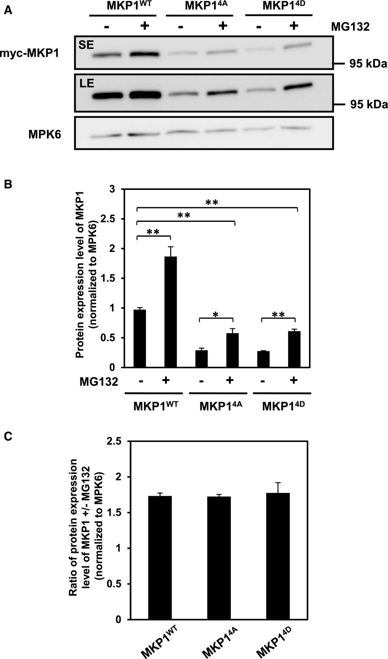
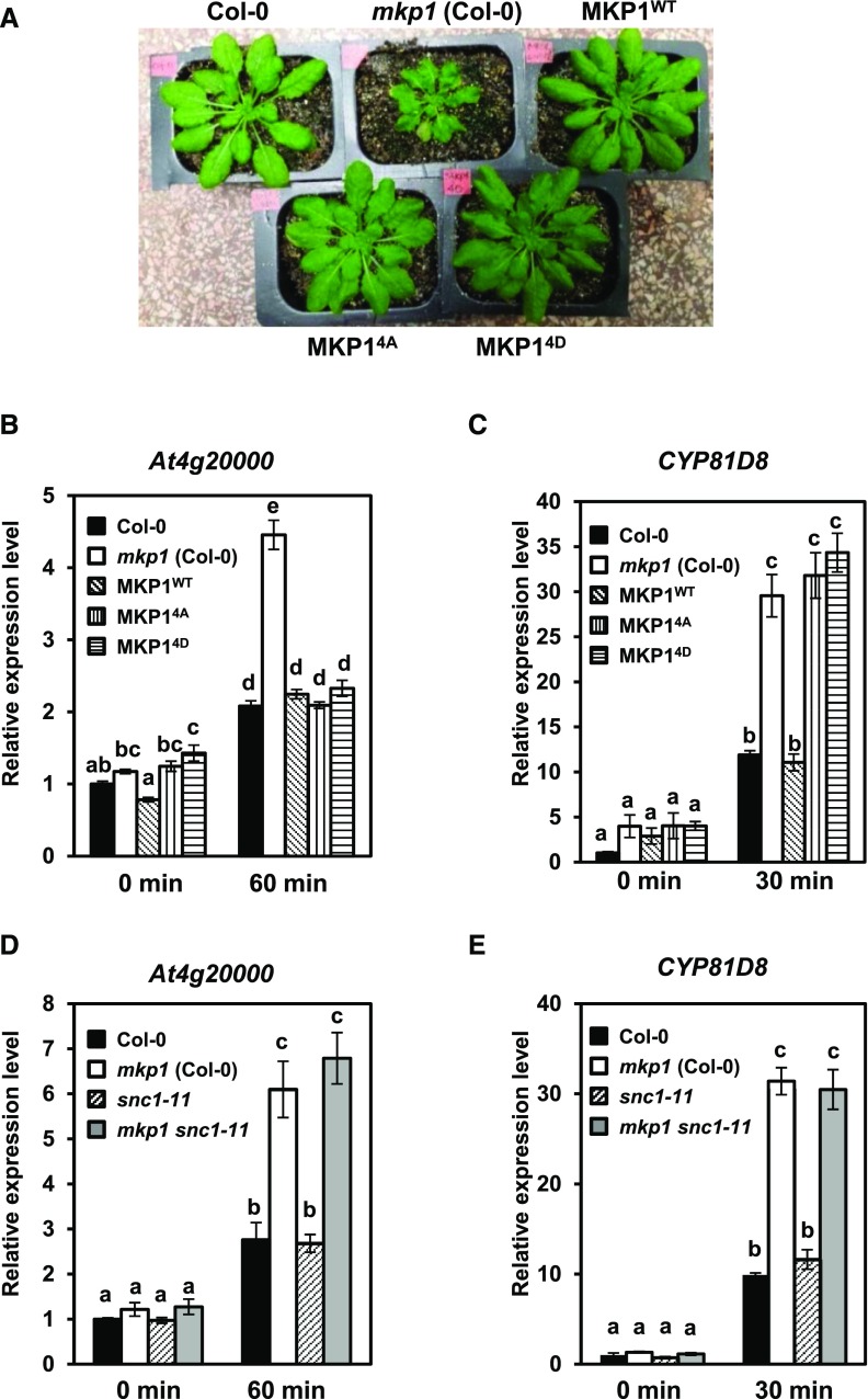
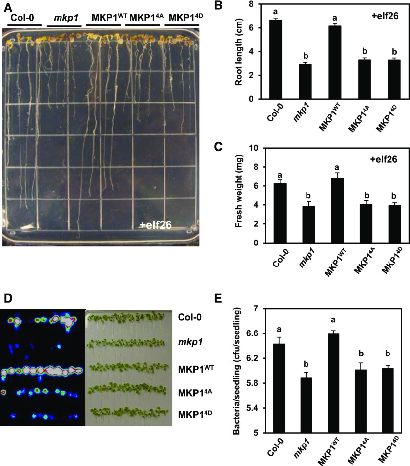
Plants perceive potential pathogens via the recognition of pathogen-associated molecular patterns (PAMPs) by surface-localized pattern recognition receptors, which initiates a series of intracellular responses that ultimately limit bacterial growth. PAMP responses include changes in intracellular protein phosphorylation, including the activation of mitogen-activated protein kinase (MAPK) cascades. MAP kinase phosphatases (MKPs), such as Arabidopsis (Arabidopsis thaliana) MKP1, are important negative regulators of MAPKs and play a crucial role in controlling the intensity and duration of MAPK activation during innate immune signaling. As such, the mkp1 mutant lacking MKP1 displays enhanced PAMP responses and resistance against the virulent bacterium Pseudomonas syringae pv tomato DC3000. Previous in vitro studies showed that MKP1 can be phosphorylated and activated by MPK6, suggesting that phosphorylation may be an important mechanism for regulating MKP1. We found that MKP1 was phosphorylated during PAMP elicitation and that phosphorylation stabilized the protein, resulting in protein accumulation after elicitation. MKP1 also can be stabilized by the proteasome inhibitor MG132, suggesting that MKP1 is constitutively degraded through the proteasome in the resting state. In addition, we investigated the role of MKP1 posttranslational regulation in plant defense by testing whether phenotypes of the mkp1 Arabidopsis mutant could be complemented by expressing phosphorylation site mutations of MKP1. The phosphorylation of MKP1 was found to be required for some, but not all, of MKP1's functions in PAMP responses and defense against bacteria. Together, our results provide insight into the roles of phosphorylation in the regulation of MKP1 during PAMP signaling and resistance to bacteria.


Figures

Figure 1.
Figure 1.
MKP1 is phosphorylated in response to elf26 in vivo. A, Fourteen-day-old seedlings expressing a myc-tagged MKP1 expressed from the CaMV 35S promoter in mkp1 (Col-0) were treated with 1 µm elf26 for the times indicated. Protein extracts from treated seedlings were separated by 8% large-format (16 × 16 cm) SDS-PAGE overnight and immunoblotted with anti-myc antibody to detect myc-MKP1 (top). The membrane was stained with Coomassie Blue (CBB) as a loading control (bottom). The stippled line aligns the faster migrating forms in the untreated control lanes on each side of the loading. B, Immunoblot analysis with anti-myc antibody of protein extracts from 14-d-old transgenic seedlings treated for 20 min with 1 µm elf26. Extracts were treated with (+) or without (−) λ-phosphatase with or without the phosphatase inhibitors. Protein extracts were separated by 8% large-format (16 × 16 cm) SDS-PAGE overnight. Experiments were performed three times with similar results to those shown.
Figure 2.
Figure 2.
MKP1 is phosphorylated by elf26-activated MPK6 on Thr-109 in vitro, but loss of MPK6 does not completely prevent elf26-induced phosphorylation in vivo. A, MPK6 was immunoprecipitated from Arabidopsis cells before and after elicitation with 100 nm elf26. The top gel is an autoradiograph of 32P incorporated in MBP, recombinant 6×His-tagged MKP1 1-161, MKP1 1-161 (T109A), or MKP1 1-161 (T64A). The bottom gel is a duplicate gel stained with Coomassie Blue (CBB) to verify equal loading. The asterisk indicates likely partially degraded MKP1. B, Pyo-MKP1 was transiently expressed from the CaMV 35S promoter in Arabidopsis protoplasts isolated from 5-week-old mkp1 (Ws) and mkp1 mpk6 (Ws) adult plants. The protoplasts were treated with 1 µm elf26 for the indicated times. Protein extracts were separated with 8% mini-format (8.3 × 7.3 cm) SDS-PAGE and immunoblotted with anti-Glu-Glu antibody to detect the Pyo-MKP1 protein (top) or the anti-phospho-p42/44 MAPKs to detect activated MAPKs (middle). The membrane was stained with Coomassie Blue as a loading control (bottom). Mock control samples were from protoplasts isolated from mkp1 (Ws) without transfection with Pyo-MKP1 plasmid. Experiments were performed at least three times with similar results to those shown.
Figure 3.
Figure 3.
Phosphorylation of MKP1 stabilizes the protein. A, Domain structure of the Arabidopsis MKP1 protein, showing the positions of the conserved putative MAPK phosphorylation sites. Underlined residues have been experimentally shown to be phosphorylated (see refs. in the text). B, Immunoblot analysis with anti-myc antibody of protein extracts from 14-d-old myc-tagged MKP1 (MKP1WT) or phosphorylation site mutant (MKP14A) transgenic seedlings treated with or without 1 µm elf26 for 20 min. Protein extracts were separated with 8% large-format (16 × 16 cm) SDS-PAGE. LE, Long exposure; SE, short exposure. C, Fourteen-day-old myc-tagged MKP1 transgenic seedlings were treated with 1 μm elf26 for the times indicated. Protein extracts from treated seedlings were separated by 8% mini-format (8.3 × 7.3 cm) SDS-PAGE and immunoblotted with anti-myc antibody to detect myc-MKP1 (top) or anti-phospho-p42/44 MAPK antibody to detect phosphorylated MAPKs (middle). Ponceau S staining of the membrane was used as a loading control (bottom). D, Fourteen-day-old transgenic seedlings expressing MKP1 wild-type protein (MKP1WT) or phosphorylation site substitutions (MKP14A and MKP14D) were treated with or without 1 µm elf26 for the times indicated. Protein extracts from treated seedlings were separated by 8% mini-format (8.3 × 7.3 cm) SDS-PAGE and immunoblotted with anti-myc antibody to detect the myc-MKP1 protein (top) or anti-MPK6 antibody as a loading control (bottom). This experiment was performed three times with similar results to those shown. E, Quantification of the western-blot band intensity of MKP1 protein normalized to the intensity of MKP6 used as a loading control. Graphed are means ± se, representative of three independent biological replicates (n = 3). Lowercase letters indicate significant groupings (P < 0.05). The statistical test was performed using ANOVA with Tukey’s pairwise comparison.
Figure 4.
Figure 4.
A portion of MKP1 is constitutively degraded by the proteasome in naïve plants. A, Fourteen-day-old transgenic seedlings expressing myc-MKP1 were treated with or without 40 µm MG132 for the times indicated. Protein extracts from treated seedlings were separated by 8% mini-format (8.3 × 7.3 cm) SDS-PAGE and immunoblotted with anti-myc antibody to detect the myc-MKP1 protein (top) or anti-MPK6 antibody used as a loading control (bottom). B, Fourteen-day-old transgenic seedlings were pretreated with or without 40 µm MG132 for 40 min and then treated with 1 µm elf26 for 20 min. Protein extracts from treated seedlings were separated by 8% mini-format (8.3 × 7.3 cm) SDS-PAGE and immunoblotted with anti-myc antibody to detect the myc-MKP1 protein (top), anti-phospho-p42/44 antibody to detect phosphorylated MAPKs (middle), or anti-MPK6 antibody as a loading control (bottom). This experiment was performed three times with similar results to those shown.
Figure 5.
Figure 5.
Phosphorylation of MKP1 is not required for MG132 stabilization. A, Fourteen-day-old transgenic seedlings were treated with or without 40 µm MG132 for 1 h. Protein extracts from treated seedlings were separated with 8% mini-format (8.3 × 7.3 cm) SDS-PAGE and immunoblotting with anti-myc antibody to detect the myc-MKP1 protein (top) and anti-MPK6 antibody as a loading control (bottom). LE, Long exposure; SE, short exposure. B and C, Quantification of the western-blot band intensity of MKP1 protein, which was normalized to that of MKP6 as a loading control. Graphed are means ± se, pooling from three independent biological replicates (n = 3). The asterisks indicate significant differences between pairwise groups (marked by parentheses: *, P < 0.05 and **, P < 0.01). The statistical test was performed using ANOVA with multiple pairwise comparisons under the protection of an overall F test.
Figure 6.
Figure 6.
MKP1 protein lacking phosphorylation sites complements some of the mkp1(Col-0) phenotypes. A, Constitutive defense-related phenotypes of adult mkp1(Col-0) plants compared with wild-type (Col-0) and transgenic mkp1 (Col-0) plants expressing myc-tagged wild-type MKP1 or phosphorylation site mutants MKP14A and MKP14D. The photograph shows 5-week-old adult plants grown on soil. B to E, mRNA levels of PAMP-responsive transcripts of At4g2000 (B and D) or CYP81D8 (C and E) measured by quantitative real-time PCR from 12-d-old seedlings treated with and without 1 μm elf26 for the indicated times. Experiments were performed to test for the requirements of MKP1 phosphorylation (B and C) or for the presence of SNC1 (D and E) in altering the accumulation pattern in mkp1 (Col-0). Transcript levels were normalized to the amount of At2g28390 transcript detected in each sample, then to the transcript level at time 0 in Col-0. Data were pooled from three independent biological experiments with an additional technical replicate for each sample (n = 6). Lowercase letters indicate significant groupings (P < 0.01). The statistical test was performed using ANOVA with Tukey’s pairwise comparison.
Figure 7.
Figure 7.
Phosphorylation of MKP1 is required for regulating elf26-induced growth inhibition and resistance to bacteria. A, Six-old-day seedlings of Col-0, mkp1 (Col-0), MKP1WT, MKP14A, and MKP14D were aseptically transferred from MS agar to the wells of a 24-well microtiter plate containing 1 mL of liquid MS medium with 1 µm elf26 for 14 d. After 14 d, seedlings were placed on an agar surface to photograph the plants, and the image is representative of at least three independent experiments. B and C, Primary root length and fresh weight were measured for the experiment described in A. Graphed are means ± se (n = 24), pooled from three independent experiments. Lowercase letters indicate significant groupings (P < 0.05). The statistical test was performed using ANOVA with Tukey’s pairwise comparison. D, Fourteen-day-old seedlings of Col-0, mkp1 (Col-0), MKP1WT, MKP14A, and MKP14D were immersed in 1 × 10⁷ cfu mL−1 DC3000 LuxCDABE. Three days post infection, seedlings were removed, rinsed with water, and placed on an agar surface. A heat map image of bacterial luminescence in DC3000-infected seedlings detected using a photon-detection camera (left) and a bright-field image of the same seedlings (right) are shown. Results are representative of three independent experiments. E, Bacterial levels in DC3000-infected seedlings 3 d post infection were measured by serial dilution plating of seedling extracts. Graphed are means ± se (n = 6). Lowercase letters indicate significant groupings (P < 0.05). The statistical test was performed using ANOVA with Tukey’s pairwise comparison. Experiments were performed three times with similar results to those shown.

Similar articles

Cited by

References

    1. Anderson JC, Bartels S, González Besteiro MA, Shahollari B, Ulm R, Peck SC (2011) Arabidopsis MAP Kinase Phosphatase 1 (AtMKP1) negatively regulates MPK6-mediated PAMP responses and resistance against bacteria. Plant J 67: 258–268 - PubMed
    1. Anderson JC, Wan Y, Kim YM, Pasa-Tolic L, Metz TO, Peck SC (2014) Decreased abundance of type III secretion system-inducing signals in Arabidopsis mkp1 enhances resistance against Pseudomonas syringae. Proc Natl Acad Sci USA 111: 6846–6851 - PMC - PubMed
    1. Asai T, Tena G, Plotnikova J, Willmann MR, Chiu WL, Gómez-Gómez L, Boller T, Ausubel FM, Sheen J (2002) MAP kinase signalling cascade in Arabidopsis innate immunity. Nature 415: 977–983 - PubMed
    1. Bartels S, Anderson JC, González Besteiro MA, Carreri A, Hirt H, Buchala A, Métraux JP, Peck SC, Ulm R (2009) MAP KINASE PHOSPHATASE1 and PROTEIN TYROSINE PHOSPHATASE1 are repressors of salicylic acid synthesis and SNC1-mediated responses in Arabidopsis. Plant Cell 21: 2884–2897 - PMC - PubMed
    1. Bartels S, González Besteiro MA, Lang D, Ulm R (2010) Emerging functions for plant MAP kinase phosphatases. Trends Plant Sci 15: 322–329 - PubMed

MeSH terms

Substances

LinkOut - more resources