

Cell adhesion molecule-1 shedding induces apoptosis of renal epithelial cells and exacerbates human nephropathies
- PMID: 29070574
- PMCID: PMC6048447
- DOI: 10.1152/ajprenal.00385.2017
Cell adhesion molecule-1 shedding induces apoptosis of renal epithelial cells and exacerbates human nephropathies
Abstract
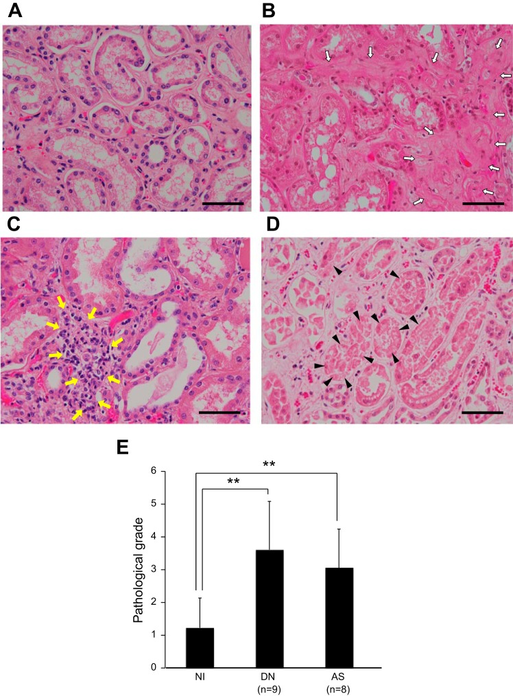
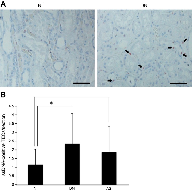
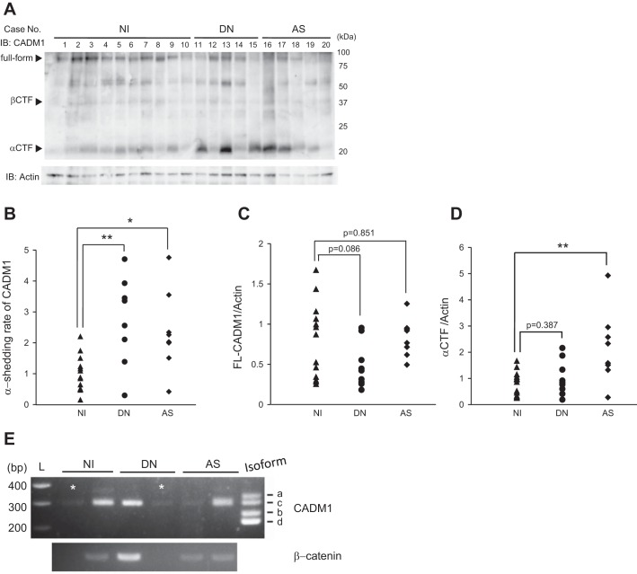
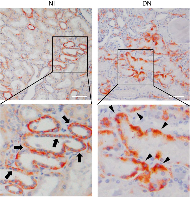
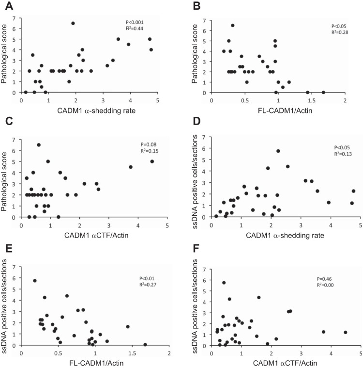
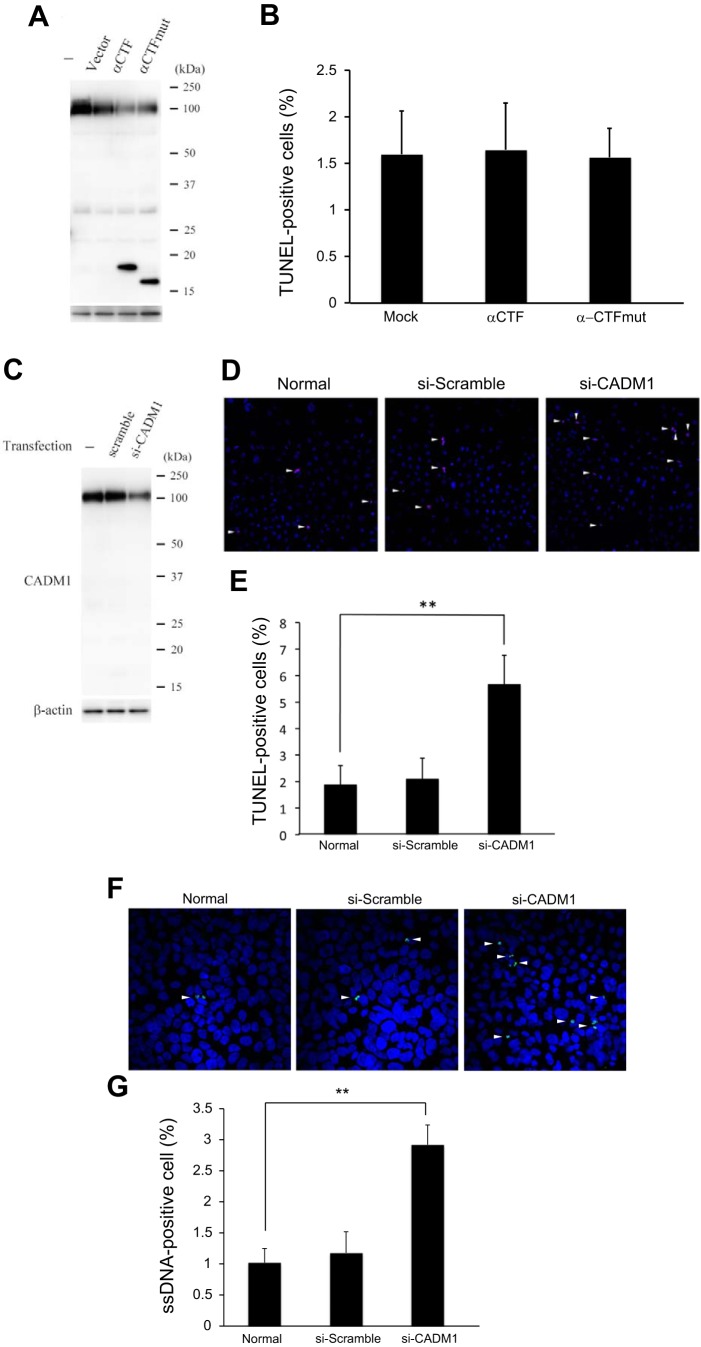
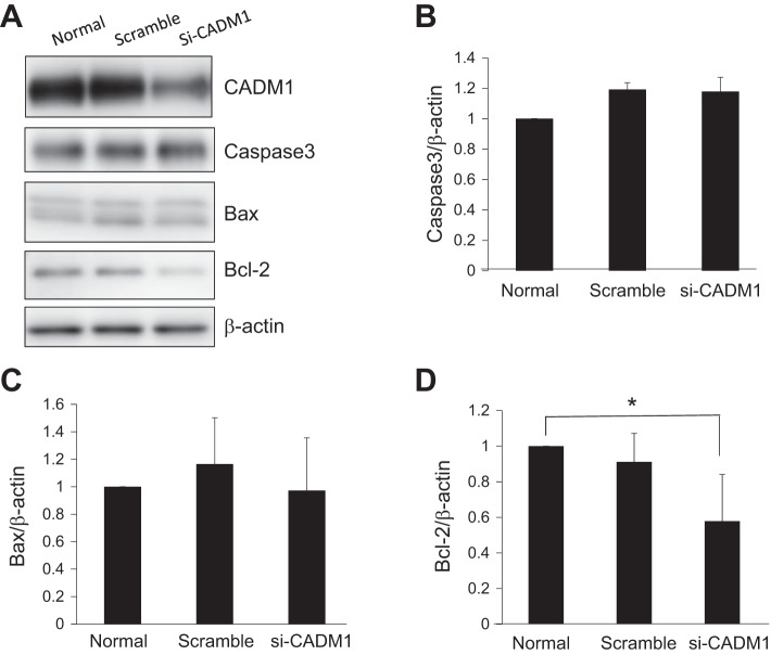
Chronic kidney disease (CKD) is an important problem throughout the world, associated with the increase of blood urea nitrogen (BUN) and serum creatinine (sCre) and with renal tubular injuries. It is crucial to elucidate the molecular mechanisms of renal injuries to identify the new therapeutics and early diagnostic methods. We focused on cell adhesion molecule-1 (CADM1) protein. CADM1, its isoform SP4, is expressed in the epithelial cells of various tissues, including renal distal tubules, localized on the lateral cell membrane, mediates cell-cell adhesion via trans-homophilic binding, and interacts with various proteins. We previously reported that its expression was downregulated by post-proteolytic cleavage (α- and β-shedding) in pulmonary diseases. To investigate whether CADM1 α-shedding occurs in human nephropathies, we performed Western blotting and immunohistochemical analysis of specimens with arterionephrosclerosis (AS) and diabetic nephropathy (DN) from autopsied kidneys. CADM1 α-shedding was induced in AS and DN kidneys and derived from the decrease in full-length CADM1 (FL-CADM1) and increase of the COOH-terminal fragment (α-CTF). In particular, the reduced FL-CADM1 level was correlated with tubular and tubulointerstitial injuries and the increases in BUN and sCre levels. Apoptosis of renal tubular epithelial cells (TECs) was promoted in both nephropathies, and it was significantly correlated with the decrease in the FL-CADM1. Furthermore, FL-CADM1 knockdown by small interfering RNA downregulated anti-apoptotic Bcl-2 protein and promoted apoptosis of cultured renal TECs. The present study suggests that the reduction of FL-CADM1 leads to renal TEC apoptosis and could exacerbate renal tubular and tubulointerstitial injuries, which contribute to the development of CKD.
Keywords: CADM1; DM nephropathy; apoptosis; arterionephrosclerotic nephropathy; shedding.
Figures
Similar articles
-
The decreased expression of electron transfer flavoprotein β is associated with tubular cell apoptosis in diabetic nephropathy.Int J Mol Med. 2016 May;37(5):1290-8. doi: 10.3892/ijmm.2016.2533. Epub 2016 Mar 18. Int J Mol Med. 2016. PMID: 27035869 Free PMC article.
-
Increased ectodomain shedding of cell adhesion molecule 1 from pancreatic islets in type 2 diabetic pancreata: correlation with hemoglobin A1c levels.PLoS One. 2014 Jun 25;9(6):e100988. doi: 10.1371/journal.pone.0100988. eCollection 2014. PLoS One. 2014. PMID: 24964098 Free PMC article.
-
Increased ectodomain shedding of cell adhesion molecule 1 as a cause of type II alveolar epithelial cell apoptosis in patients with idiopathic interstitial pneumonia.Respir Res. 2015 Aug 1;16:90. doi: 10.1186/s12931-015-0255-x. Respir Res. 2015. PMID: 26231557 Free PMC article.
-
Multiple Functions of Cell Adhesion Molecule 1 (CADM1) and Its Role in the Pathogenesis of Cancer and Other Diseases.J Nippon Med Sch. 2025;92(2):122-131. doi: 10.1272/jnms.JNMS.2025_92-205. J Nippon Med Sch. 2025. PMID: 40399107 Review.
-
Proteinuria and tubular cells: Plasticity and toxicity.Acta Physiol (Oxf). 2025 Feb;241(2):e14263. doi: 10.1111/apha.14263. Acta Physiol (Oxf). 2025. PMID: 39797499 Review.
Cited by
-
Brucella melitensis UGPase inhibits the activation of NF-κB by modulating the ubiquitination of NEMO.BMC Vet Res. 2021 Aug 30;17(1):289. doi: 10.1186/s12917-021-02993-9. BMC Vet Res. 2021. PMID: 34461896 Free PMC article.
-
Dezocine inhibits cell proliferation, migration, and invasion by targeting CRABP2 in ovarian cancer.Open Med (Wars). 2022 Dec 14;17(1):2052-2061. doi: 10.1515/med-2022-0541. eCollection 2022. Open Med (Wars). 2022. PMID: 36568517 Free PMC article.
-
ADAM10 and ADAM17, Major Regulators of Chronic Kidney Disease Induced Atherosclerosis?Int J Mol Sci. 2023 Apr 15;24(8):7309. doi: 10.3390/ijms24087309. Int J Mol Sci. 2023. PMID: 37108478 Free PMC article. Review.
-
Cell Adhesion Molecule 1 Contributes to Cell Survival in Crowded Epithelial Monolayers.Int J Mol Sci. 2020 Jun 9;21(11):4123. doi: 10.3390/ijms21114123. Int J Mol Sci. 2020. PMID: 32527032 Free PMC article.
-
Apoptosis in kidney tissue of senior and geriatric cats with chronic kidney disease.J Vet Med Sci. 2025 Mar 1;87(3):248-256. doi: 10.1292/jvms.24-0296. Epub 2025 Jan 22. J Vet Med Sci. 2025. PMID: 39842786 Free PMC article.
References
-
- Atapattu L, Saha N, Chheang C, Eissman MF, Xu K, Vail ME, Hii L, Llerena C, Liu Z, Horvay K, Abud HE, Kusebauch U, Moritz RL, Ding BS, Cao Z, Rafii S, Ernst M, Scott AM, Nikolov DB, Lackmann M, Janes PW. An activated form of ADAM10 is tumor selective and regulates cancer stem-like cells and tumor growth. J Exp Med 213: 1741–1757, 2016. doi:10.1084/jem.20151095. - DOI - PMC - PubMed
-
- Athyros VG, Tziomalos K, Katsiki N, Doumas M, Karagiannis A, Mikhailidis DP. Cardiovascular risk across the histological spectrum and the clinical manifestations of non-alcoholic fatty liver disease: An update. World J Gastroenterol 21: 6820–6834, 2015. doi:10.3748/wjg.v21.i22.6820. - DOI - PMC - PubMed
-
- Colombini M. Pore size and properties of channels from mitochondria isolated from Neurospora crass. J Membr Biol 53: 79–84, 1980. doi:10.1007/BF01870576. - DOI
Publication types
MeSH terms
Substances
LinkOut - more resources
Full Text Sources
Other Literature Sources
Medical
Research Materials
Miscellaneous