. 2017 Nov 21;89(21):2157-2166.
doi: 10.1212/WNL.0000000000004669. Epub 2017 Oct 25.

Multiparametric MRI changes persist beyond recovery in concussed adolescent hockey players

Affiliations

Multiparametric MRI changes persist beyond recovery in concussed adolescent hockey players

Kathryn Y Manning et al. Neurology. .

Erratum in

Abstract

Objective: To determine whether multiparametric MRI data can provide insight into the acute and long-lasting neuronal sequelae after a concussion in adolescent athletes.

Methods: Players were recruited from Bantam hockey leagues in which body checking is first introduced (male, age 11-14 years). Clinical measures, diffusion metrics, resting-state network and region-to-region functional connectivity patterns, and magnetic resonance spectroscopy absolute metabolite concentrations were analyzed from an independent, age-matched control group of hockey players (n = 26) and longitudinally in concussed athletes within 24 to 72 hours (n = 17) and 3 months (n = 14) after a diagnosed concussion.

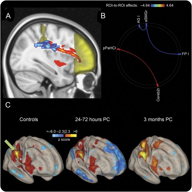
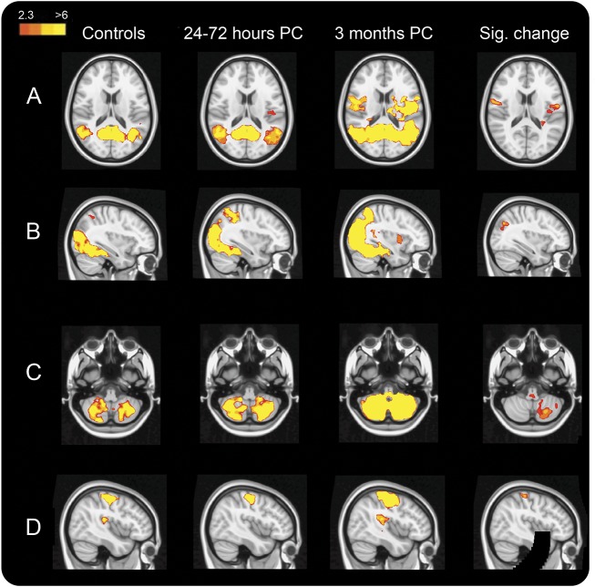
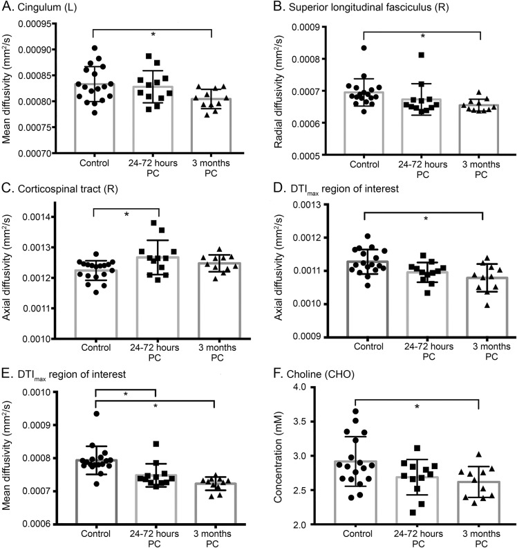
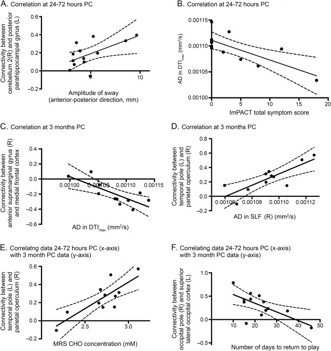
Results: There were diffusion abnormalities within multiple white matter tracts, functional hyperconnectivity, and decreases in choline 3 months after concussion. Tract-specific spatial statistics revealed a large region along the superior longitudinal fasciculus with the largest decreases in diffusivity measures, which significantly correlated with clinical deficits. This region also spatially intersected with probabilistic tracts connecting cortical regions where we found acute functional connectivity changes. Hyperconnectivity patterns at 3 months after concussion were present only in players with relatively less severe clinical outcomes, higher choline concentrations, and diffusivity indicative of relatively less axonal disruption.

Conclusions: Changes persisted well after players' clinical scores had returned to normal and they had been cleared to return to play. Ongoing white matter maturation may make adolescent athletes particularly vulnerable to brain injury, and they may require extended recovery periods. The consequences of early brain injury for ongoing brain development and risk of more serious conditions such as second impact syndrome or neural degenerative processes need to be elucidated.


Figures

Figure 1
Figure 1. Acute connectivity changes spatially relate to axonal damage
(A) Frontal pole (FP) and anterior supramarginal gyrus (aSMG) are shown in yellow, with the probabilistic tractography connecting them shown in red. DTImax region is overlaid in blue, showing the spatial relationship between structural abnormalities and resting-state fMRI connectivity. (B) Significant whole-brain region-to-region connectivity differences between controls and the 24- to 72-hour postconcussion (PC) group. Blue indicates a decrease and red indicates an increase in region-to-region connectivity. (C) Voxel-wise connectivity pattern using the right aSMG (green arrow) as a seed, displaying the enhanced anticorrelation (blue) with the FP and regions that mimic the default mode network. AG = angular gyrus; pPaHC = posterior parahippocampus; ROI = region of interest.
Figure 2
Figure 2. RSN hyperconnectivity at 3 months after concussion
Each column represents the average resting-state network (RSN) for each of the 3 groups (scaled by z statistic), with areas that are significantly more highly correlated with that network at 3 months compared to (A–C) controls or (D) the 24- to 72-hour postconcussion (PC) group (p < 0.001 except for [A] where p < 0.01). Results are shown for the (A) default mode network, (B) occipital pole visual RSN, (C) cerebellar RSN, and (D) sensorimotor RSN.
Figure 3
Figure 3. Region-to-region hyperconnectivity at 3 months
Significantly increased connectivity (red lines) is shown using (A) connectome rings with regions labeled around the perimeter and (B) 3-dimensional brain volumes in which the color of the spheres and the transparency of the connecting lines indicate the strength of the effect using a t statistic after false discovery rate correction (corrected p < 0.05). aSMG = anterior supramarginal gyrus; Cereb = cerebellum; CO = central opercular cortex; dmn.MPFC = default mode network medial prefrontal cortex; HG = Heschl gyrus; MedFC = medial frontal cortex; MidFG = middle frontal gyrus; OFusG = occipital fusiform gyrus; OP = occipital pole; PaCiG = paracingulate gyrus; pMTG = posterior middle temporal gyrus; PO = parietal operculum cortex; Pre/PostCG = precentral/postcentral gyri; sLOC = superior lateral occipital cortex; SPL = superior parietal lobule; toITG = temporoccipital inferior temporal gyrus; TP = temporal pole; Ver = vermis.
Figure 4
Figure 4. Diffusion and MRS results
(A–E) Average diffusion metrics within standard atlas-derived white matter tracts and (F) absolute metabolite concentrations with the prefrontal white matter voxel. SDs are shown with error bars. *Significant multivariate analysis of variance–corrected post hoc differences (p < 0.05). DTI = diffusion tensor imaging; MRS = magnetic resonance spectroscopy; PC = postconcussion.
Figure 5
Figure 5. Examples of correlation results
Relating data at (A and B) 24 to 72 hours postconcussion (PC), (C and D) 3 months PC, and (E and F) between 24 to 72 hours PC (x-axis) and 3 months PC (y-axis) data. Further details are provided in table e-2. The 95% confidence interval is shown using the dashed curves, and all relationships shown here have p < 0.05 after false discovery rate correction. AD = axial diffusivity; CHO = choline; DTI = diffusion tensor imaging; ImPACT = Immediate Post-Concussion Assessment and Cognitive Testing; MRS = magnetic resonance spectroscopy; SLF = superior longitudinal fasciculus.

References

    1. Benz B, Ritz A, Kiesow S. Influence of age-related factors on long-term outcome after traumatic brain injury (TBI) in children: a review of recent literature and some preliminary findings. Restor Neurol Neurosci 1999;14:135–141. - PubMed
    1. Cusimano MD, Cho N, Amin K, et al. Mechanisms of team-sport-related brain injuries in children 5 to 19 years old: opportunities for prevention. PLoS One 2013;8:e58868. - PMC - PubMed
    1. Bigler ED, Maxwell WL. Neuropathology of mild traumatic brain injury: relationship to neuroimaging findings. Brain Imaging Behav 2012;6:108–136. - PubMed
    1. Borich M, Babul AN, Yuan PH, Boyd L, Virji-Babul N. Alterations in resting-state brain networks in concussed adolescent athletes. J Neurotrauma 2015;32:265–271. - PubMed
    1. Mayer AR, Ling JM, Yang Z, Pena A, Yeo RA, Klimaj S. Diffusion abnormalities in pediatric mild traumatic brain injury. J Neurosci 2012;32:17961–17969. - PMC - PubMed