. 2017 Dec 1;199(11):3849-3857.
doi: 10.4049/jimmunol.1601540. Epub 2017 Oct 25.

IL-17A-Induced PLET1 Expression Contributes to Tissue Repair and Colon Tumorigenesis

Affiliations

IL-17A-Induced PLET1 Expression Contributes to Tissue Repair and Colon Tumorigenesis

Jarod A Zepp et al. J Immunol. .

Abstract

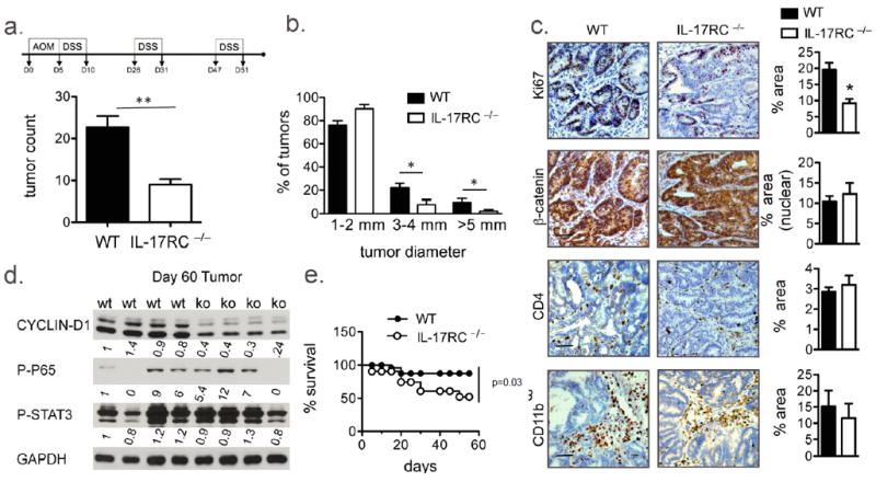
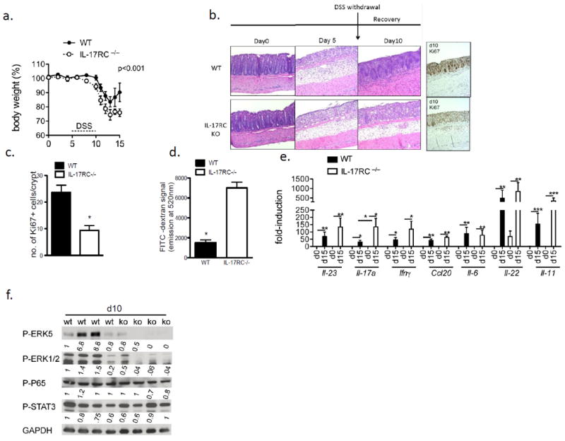
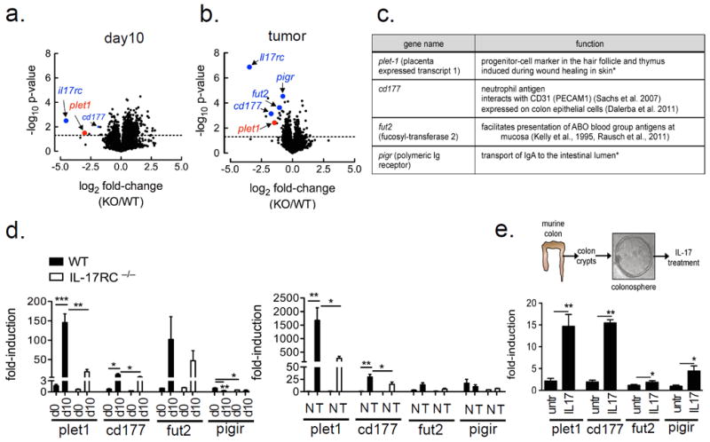
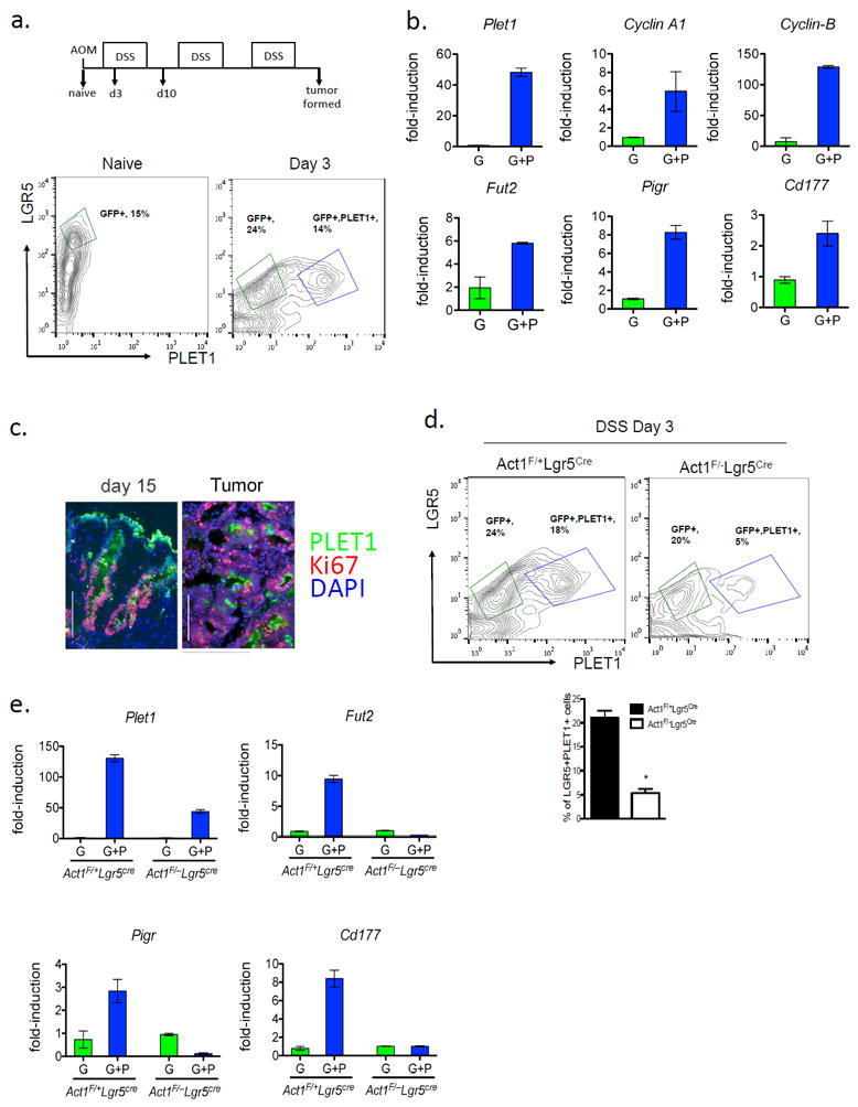
This study identifies a novel mechanism linking IL-17A with colon tissue repair and tumor development. Abrogation of IL-17A signaling in mice attenuated tissue repair of dextran sulfate sodium (DSS)-induced damage in colon epithelium and markedly reduced tumor development in an azoxymethane/DSS model of colitis-associated cancer. A novel IL-17A target gene, PLET1 (a progenitor cell marker involved in wound healing), was highly induced in DSS-treated colon tissues and tumors in an IL-17RC-dependent manner. PLET1 expression was induced in LGR5+ colon epithelial cells after DSS treatment. LGR5+PLET1+ marks a highly proliferative cell population with enhanced expression of IL-17A target genes. PLET1 deficiency impaired tissue repair of DSS-induced damage in colon epithelium and reduced tumor formation in an azoxymethane/DSS model of colitis-associated cancer. Our results suggest that IL-17A-induced PLET1 expression contributes to tissue repair and colon tumorigenesis.


Conflict of interest statement

Conflict of Interests

The authors declare no conflict of interest.

Figures

Figure 1
Figure 1. IL-17RC mediates colon tumorigenesis in CAC model
(a, top) Schematic of CAC model (a) Tumor number (b) Tumor size distribution (a and b n=12 per group), (c) IHC images from WT and IL-17RC-deficient tumor tissue stained for the indicated markers. (d) Immunoblot analysis of colon tumor tissue from the indicated mice, each lane represents one mouse. (e) Percent survival of the indicated mice throughout the CAC model. Representative images and data are shown in (c). Quantification was performed on staining of 5 tumors from each group and in 3 views per sample. Error bars represent mean ± SEM scale bars =100μm, p values shown are * <0.05.
Figure 2
Figure 2. IL-17RC-deficiency impacts colon epithelial integrity and wound healing response
(a) Weight loss during first 15 days of CAC (b) H&E or IHC staining for Ki67 in colon tissue from DSS-treated WT and IL-17RC-deficient mice. Mice were treated with DSS for 5-days followed by 5 days of regular water to allow for the epithelium to recover. (c) Quantification of Ki67 staining. (d) Serum FITC signal from mice treated with 3.5% DSS for 5 days followed by DSS withdrawal for 5 days, and then gavaged with FITC-dextran. Data shown is from a representative experiment with n=4 mice per group. (e) RT-qPCR from colon tissue of mice from day 10 of the DSS treatment, n=4 mice per group. All gene expression values were normalized to β-actin. (f) Immunoblot analysis of DSS treated colon tissue from WT or IL-17RC-deficient (KO) mice. Each lane represents a single mouse. Quantification was performed on staining of 3 colon sections from each group and in 3 views per sample. Experiments were reproduced 3 times, error bars represent mean ± SEM scale bars =100μm, p values shown are * <0.05, ** <0.01, *** <0.001.
Figure 3
Figure 3. IL-17RC-dependent gene analysis during regeneration and in established tumors
Volcano plots from Affymetrix gene-array comparing (a) day 10 (3.5% DSS for 5 days followed by DSS withdrawal for 5 days) colon tissue (n=4 per group) and (b) tumor tissue (n=5-6 per group) mRNA between WT and IL-17RC-deficient mice. Expression data were averaged and p-values derived by fold-change in expression between IL-17RC-deficient and WT, dotted line represents a p-value of 0.05 as determined by unpaired two-tailed t-test. (c) Table of genes significantly reduced in the tumors of IL-17RC-deficient mice, and their known functions, * references as cited in the text. (d) Gene expression by RT-qPCR from untreated and DSS Day 10 treated colon tissue, n=5 mice per group or (e) RT-qPCR results from normal-adjacent (N) and tumor tissue (T), n=5-6 mice per group. (f, top) Schematic of colonosphere isolation and IL-17A treatment procedure. (f, bottom) Gene expression by RT-qPCR from colonospheres generated from WT mice. Spheres were grown for one week and then treated with IL-17A (50 ng/ml) for 24 hours, shown are representative data from 3 independent experiments, RT-qPCR data in d-f were normalized to β-actin. Data presented as mean ± SEM, p values shown are * <0.05, ** <0.01, ***<0.001.
Figure 4
Figure 4. PLET1 is induced from colonic crypts during DSS treatment
(a) FACS analysis of colonic epithelial cells derived from DSS treated Lgr5EGFP-IRES-CreERT2 mice, n=3 mice combined for each treatment group. (b) Gene expression from FACS-sorted cell populations from day 3 of DSS treatment; G, LGR5-eGFP-positive, GP, LGR5-eGFP/PLET1-positive, all RT-qPCR results are relative to Gapdh. (c) Immunofluorescence staining of PLET1 and Ki67 in colon tissue and tumors from AOM-DSS treated mice. (d) Representative flow cytometry analysis of isolated colonic epithelial cells and quantification of FACS data from the indicated mice, n=3 mice combined for each group. (e) RT-qPCR from the indicated FACS sorted cell populations relative to Gapdh. Data are representative of 2 independent experiments are presented as mean ± SEM.
Figure 5
Figure 5. PLET1 is an ERK5-target and promotes cell transformation and P-ERK1/2 signaling
(a) Lysates from colon crypts of WT mice untreated or stimulated with IL-17A (50 ng/ml) were analyzed by western blotting with the indicated antibodies. (b) Serum FITC-dextran from DSS treated mice injected with BIX02189 or vehicle control, shown are representative results from two independently performed experiments. (c) Western analysis of lysates from colon epithelial cells of mice treated with BIX01289 and DSS for 5 days followed by 5 days of DSS removal. Each lane is from one mouse. Band intensity normalized to β-ACTIN is indicated below the blots. (d-f) Plet1-/- (n=5) and Plet1+/- (n=6) littermate control mice were subjected to 3.5% DSS treatment for 5 days followed by 5 days of DSS withdrawal. On day 10 mice were subjected to gut permeability assay using FITC dextran (e). The mice were sacrificed and the colons were taken for H&E and Ki67 staining (d) or western blot analysis (f). (g) Plet1+/- (n=15) and Plet1-/- (n=10) mice were subjected to AOM-DSS regiment. Number of tumors per mouse and tumor size distribution were plotted. (h) Representative image of H&E and Ki67 staining of tumors from mice of indicated genotypes. Quantification is shown as bar graph to the right. Quantification of histology was performed on staining of 3 colon sections from each group and in 3 views per sample. All data presented are means ± SEM, p values shown are * <0.05.

References

    1. Kluwe J, Mencin A, Schwabe RF. Toll-like receptors, wound healing, and carcinogenesis. Journal of molecular medicine. 2009;87:125–138. - PMC - PubMed
    1. Ivanov II, Atarashi K, Manel N, Brodie EL, Shima T, Karaoz U, Wei D, Goldfarb KC, Santee CA, Lynch SV, Tanoue T, Imaoka A, Itoh K, Takeda K, Umesaki Y, Honda K, Littman DR. Induction of intestinal Th17 cells by segmented filamentous bacteria. Cell. 2009;139:485–498. - PMC - PubMed
    1. Punkenburg E, Vogler T, Buttner M, Amann K, Waldner M, Atreya R, Abendroth B, Mudter J, Merkel S, Gallmeier E, Rose-John S, Neurath MF, Hildner K. Batf-dependent Th17 cells critically regulate IL-23 driven colitis-associated colon cancer. Gut. 2016;65:1139–1150. - PubMed
    1. Chae WJ, Gibson TF, Zelterman D, Hao L, Henegariu O, Bothwell AL. Ablation of IL-17A abrogates progression of spontaneous intestinal tumorigenesis. Proceedings of the National Academy of Sciences of the United States of America. 2010;107:5540–5544. - PMC - PubMed
    1. Grivennikov SI, Wang K, Mucida D, Stewart CA, Schnabl B, Jauch D, Taniguchi K, Yu GY, Osterreicher CH, Hung KE, Datz C, Feng Y, Fearon ER, Oukka M, Tessarollo L, Coppola V, Yarovinsky F, Cheroutre H, Eckmann L, Trinchieri G, Karin M. Adenoma-linked barrier defects and microbial products drive IL-23/IL-17A-mediated tumour growth. Nature. 2012;491:254–258. - PMC - PubMed

Publication types

MeSH terms