. 2017 Oct 25;7(1):13977.
doi: 10.1038/s41598-017-13939-3.

Melanopsin, a Canonical Light Receptor, Mediates Thermal Activation of Clock Genes

Affiliations

Melanopsin, a Canonical Light Receptor, Mediates Thermal Activation of Clock Genes

Maria Nathália Moraes et al. Sci Rep. .

Abstract

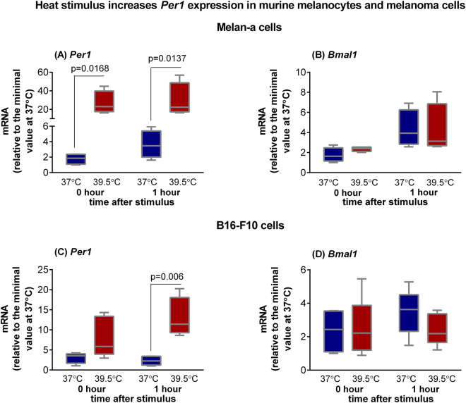
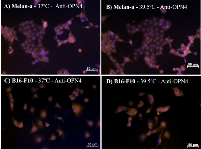
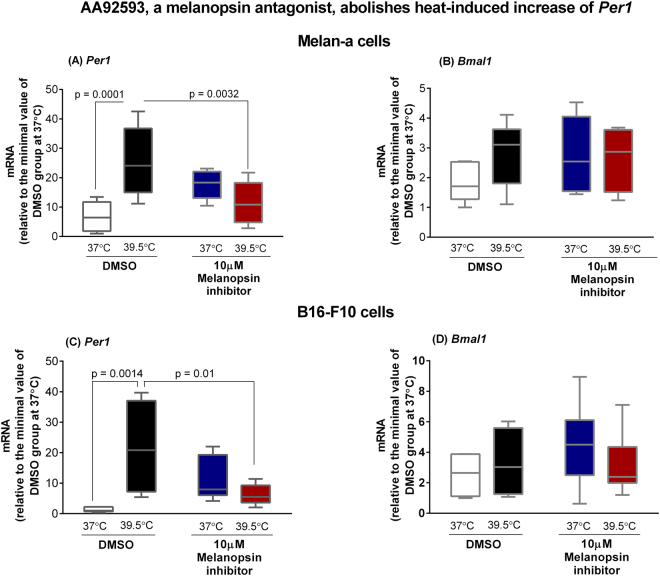
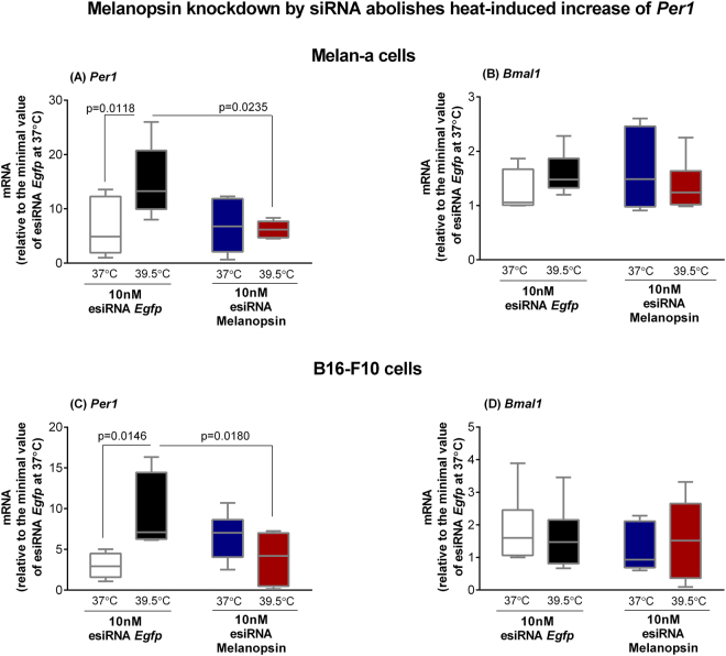
Melanopsin (OPN4) is a photo-pigment found in a small subset of intrinsically photosensitive ganglion cells (ipRGCs) of the mammalian retina. These cells play a role in synchronizing the central circadian pacemaker to the astronomical day by conveying information about ambient light to the hypothalamic suprachiasmatic nucleus, the site of the master clock. We evaluated the effect of a heat stimulus (39.5 °C) on clock gene (Per1 and Bmal1) expression in cultured murine Melan-a melanocytes synchronized by medium changes, and in B16-F10 melanoma cells, in the presence of the selective OPN4 antagonist AA92593, or after OPN4 knockdown by small interfering RNA (siRNA). In addition, we evaluated the effects of heat shock on the localization of melanopsin by immunocytochemistry. In both cell lines melanopsin was found in a region capping the nucleus and heat shock did not affect its location. The heat-induced increase of Per1 expression was inhibited when melanopsin was pharmacologically blocked by AA92593 as well as when its protein expression was suppressed by siRNA in both Melan-a and B16-F10 cells. These data strongly suggest that melanopsin is required for thermo-reception, acting as a thermo-opsin that ultimately feeds the local circadian clock in mouse melanocytes and melanoma cells.


Conflict of interest statement

The authors declare that they have no competing interests.

Figures

Figure 1
Figure 1
Expression of Per1 (A and C) and Bmal1 (B and D) in murine Melan-a melanocytes and B16-F10 melanoma cells after heat stimulus (39.5 °C). Melan-a or B16-F10 cells were kept for 3 days in constant dark and temperature (37 °C). In the beginning of the 4th day, Melan-a cells were synchronized by two medium changes and after further 24 hours they were heat-stimulated (39.5 °C) during 1 h. B16-F10 cells were heat stimulated in the beginning of the 4th day. Total RNA was extracted immediately and 1 h after the end of the stimulus for Melan-a and B16-F10 cells. Boxplots show the median, quartiles, maximum, and minimum expression values of each gene transcript normalized by 18S ribosomal RNA (for Melan-a cells) and Rpl37a (for B16-F10 cells), and expressed relative to the minimal value at 37 °C (N = 4–6). Statistical analysis was performed by Two-way ANOVA followed by Bonferroni post-test.
Figure 2
Figure 2
Representative fields of melanopsin (OPN4) immunostaining in Melan-a (A,B) and B16-F10 (C,D) cells. Cells were kept in DD for 3 days and at the beginning of the 4th day, cells were divided into 2 groups: (1) Control group kept in constant dark and temperature (37 °C); (2) group in constant dark and exposed to 1 h heat stimulus (39.5 °C). Twenty-four hours later the medium was removed and the cells were fixed with 4% paraformaldehyde. DAPI stained nuclei in blue and OPN4 immunopositivity (1:500 antiserum), revealed with a Cy3-labeled secondary antibody, in orange. Photomicrographies were taken with Axiocam MRm camera (Zeiss) and pseudocolored with Axiovision software (Zeiss). Scale bar 50 μm (200x magnification).
Figure 3
Figure 3
Expression of Per1 (A and C) and Bmal1 (B and D) in murine Melan-a melanocytes and B16-F10 melanoma cells after heat stimulus (39.5 °C) in the presence of AA92593. Melan-a or B16-F10 cells were kept for 3 days in constant dark and temperature (37 °C). In the beginning of the 4th day, Melan-a cells were synchronized by two medium changes, and after further 24 hours cells were heat-stimulated. For B16-F10 cells, the heat shock (39.5 °C) was applied at the beginning of the 4th day. In both scenarios, cells were divided into four groups: (1) control group at 37 °C in the presence of DMSO (0.1%); (2) heat-stimulated (39.5 °C) group in the presence of DMSO (0.1%); (3) group kept at 37 °C in the presence of AA92593 (10 µM), a selective OPN4 antagonist; (4) heat-stimulated (39.5 °C) group in the presence of AA92593 (10 µM). Total RNA was extracted immediately and 1 h after the end of the stimulus for Melan-a and B16-F10 cells, respectively. Boxplots show the median, quartiles, maximum, and minimum expression values of each gene transcript normalized by Rpl 37a and expressed relative to the minimal value of DMSO group at 37 °C (N = 5–9). Statistical analysis was performed by One-way ANOVA followed by Tukey’s test.
Figure 4
Figure 4
Representative fields of melanopsin (OPN4) immunostaining in Melan-a (A,B) and B16-F10 (C,D) cells. Cells were kept in DD for 24 hours, on the 2nd day, were transfected with esiRNA against melanopsin or EGFP (both at 10 nM), and 48 h after transfection the cells were immunostained for melanopsin (OPN4). (A and C) esiRNA against mRNA of EGFP transfected cells (control group) and (B and D) esiRNA against mRNA of OPN4 transfected cells. Photo-micrographies were obtained with 200 x magnification in an inverted fluorescence microscope Axiovert 40CFL (Zeiss, Oberkochen, Germany) with a mercury lamp of 50 W, and DAPI (excitation 358 and emission 463 nm) and Cy3 (excitation 549 and emission 562 nm) filters. Melanopsin gene and protein knockdown by endoribonuclease-prepared siRNAs (esiRNA). Gene expression of Opn4 (melanopsin encoding gene) in esiRNA against mRNA of EGFP (control group) or of melanopsin transfected cells. Melan-a (E) or B16-F10 (F) cells were kept during three days in constant dark and temperature (37 °C). At the beginning of the 4th day, Melan-a cells were synchronized by two medium changes, and after further 24 hours they were transfected with esiRNA. B16-F10 cells were transfected at the beginning of the 4th day. In both cases, gene expression was evaluated 48 h after transfection with esiRNA. Boxplots show the median, quartiles, maximum, and minimum expression values of each gene transcript normalized by Rpl 37a and expressed relative to the minimal value of the esiRNA EGFP group kept at 37 °C (N = 5–6). Statistical analysis was performed by Student’s t test.
Figure 5
Figure 5
Expression of Per1 (A and C) and Bmal1 (B and D) in esiRNA transfected murine Melan-a melanocytes and B16-F10 melanoma cells after heat stimulus (39.5 °C). Melan-a or B16-F10 cells were kept during three days in constant dark and temperature (37 °C). At the beginning of the 4th day, Melan-a cells were synchronized by two medium changes, and after further 24 h cells were transfected with esiRNA against melanopsin or EGFP (both at 10 nM) using Lipofectamine 3000 transfection kit. B16-F10 cells were transfected with esiRNA, as described above, at the beginning of the 4th day. In both experimental scenarios, 48 hours after transfection, cells were divided into four groups: (1) control group at 37 °C in the presence of esiRNA EGFP (10 nM); (2) heat-stimulated (39.5 °C) group in the presence of esiRNA EGFP (10 nM); (3) group at 37 °C in the presence of esiRNA melanopsin (10 nM); (4) heat-stimulated (39.5 °C) group in the presence of esiRNA melanopsin (10 nM). Boxplots show the median, quartiles, maximum, and minimum expression values of each gene transcript normalized by Rpl 37a and expressed relative to the minimal value of the esiRNA EGFP group at 37 °C (N = 5–11). Total RNA was extracted immediately and 1 h after the end of the stimulus for Melan-a and B16-F10 cells, respectively. Statistical analysis was performed by One-way ANOVA followed by Tukey’s test.

Similar articles

Cited by

References

    1. Provencio I, Rollag MD, Castrucci AML. Photoreceptive net in the mammalian retina. This mesh of cells may explain how some blind mice can still tell day from night. Nature. 2002;415(6871):493. doi: 10.1038/415493a. - DOI - PubMed
    1. Panda S, et al. Melanopsin is required for non-image-forming photic responses in blind mice. Science. 2003;301(5632):525–527. doi: 10.1126/science.1086179. - DOI - PubMed
    1. Ralph MR, Foster RG, Davis FC, Menaker M. Transplanted suprachiasmatic nucleus determines circadian period. Science. 1990;247(4945):975–978. doi: 10.1126/science.2305266. - DOI - PubMed
    1. Allen AE, Storchi R, Martial FP, Bedford RA, Lucas RJ. Melanopsin contributions to the representation of images in the early visual system. Curr. Biol. 2017;27(11):1623–1632. doi: 10.1016/j.cub.2017.04.046. - DOI - PMC - PubMed
    1. Hughes S, et al. Signalling by melanopsin (OPN4) expressing photosensitive retinal ganglion cells. Eye (Lond) 2016;30(2):247–254. doi: 10.1038/eye.2015.264. - DOI - PMC - PubMed

Publication types