. 2017 Oct 25;7(1):14048.
doi: 10.1038/s41598-017-12968-2.

Dual negative roles of C/EBPα in the expansion and pro-tumor functions of MDSCs

Affiliations

Dual negative roles of C/EBPα in the expansion and pro-tumor functions of MDSCs

John R Mackert et al. Sci Rep. .

Abstract

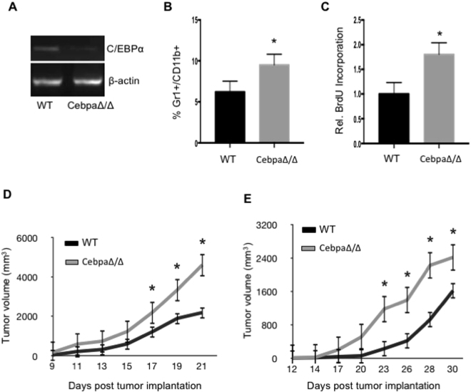
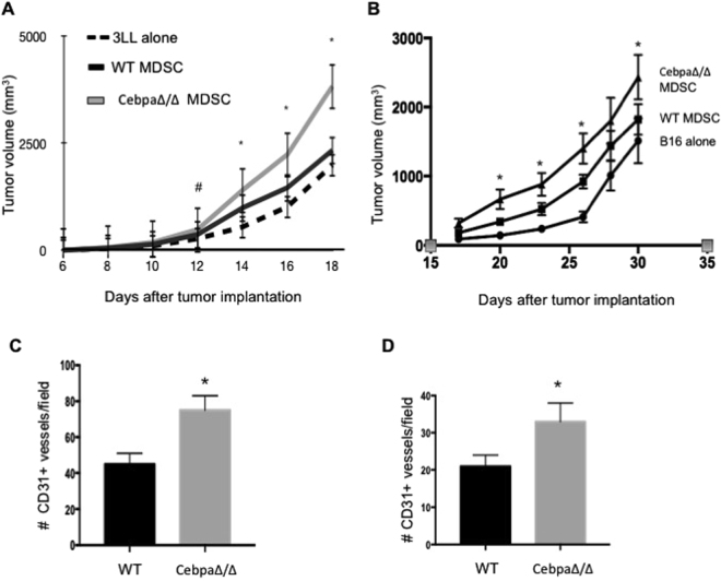
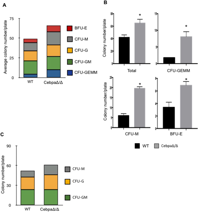
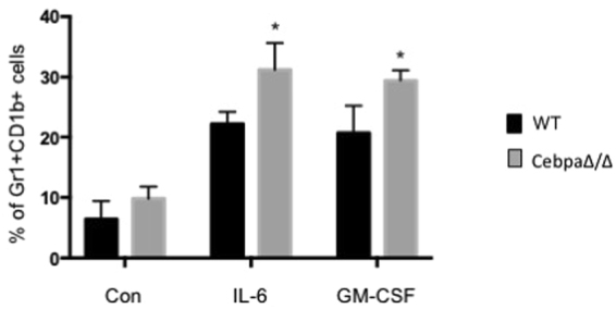
Myeloid-derived suppressor cells (MDSCs) are greatly expanded in cancer patients and tumor-bearing mice. They infiltrate into tumors and modulate the tumor microenvironment. In an effort to identify molecular mediators responsible for expansion and the tumor-promoting function of MDSCs, we discovered CCAAT/enhancer binding protein alpha (C/EBPα) expression was significantly reduced in MDSCs from tumor-bearing mice compared to non-tumor-bearing hosts. Tumor-conditioned medium down-regulated C/EBPα expression, suggesting tumor secreted factors inhibiting the gene expression. Consistent with the function of C/EBPα in regulating the balance between proliferation and growth arrest in hematopoietic progenitors, myeloid lineage specific deletion of C/EBPα resulted in significantly enhanced MDSC proliferation and expansion, as well as an increase of myeloid progenitors and a decrease of mature cells. In addition, deletion of C/EBPα in MDSCs enhanced the pro-angiogenic, immune suppressive and pro-tumorigenic behavior of these cells by upregulating the production of iNOS and arginase, as well as MMP-9 and VEGF. Accordingly, tumors growing in C/EBPα conditional null mice displayed greater MDSC infiltration, increased vascularization and accelerated tumor growth. Taken together, this study reveals dual negative roles of C/EBPα in the expansion as well as pro-angiogenic and immune suppressive functions in MDSCs.


Conflict of interest statement

The authors declare that they have no competing interests.

Figures

Figure 1
Figure 1
C/EBPα is down-regulated in MDSCs from tumor bearing mice. Gr-1+CD11b+ cells were isolated from spleens (spln) of C57BL/6 mice with (+) or without 3LL (−) tumors as well as tumor tissues (tumor). Gr-1+CD11b+ cells with greater than 95% purity were pooled from 5–7 mice, RNA was isolated and C/EBPα expression was measured by real-time PCR (A). Media was collected from 3LL tumor cells after 2–3 days in culture to make tumor-conditioned medium (TCM). 32D cells were cultured in a 50:50 mixture of TCM and fresh media for 4 or 48 hours. RNA was isolated and C/EBPα expression was measured by real-time PCR (B). *p < 0.05. The data are presented as mean with SD. The experiment was done in triplicate and repeated twice.
Figure 2
Figure 2
Tumor growth is accelerated in CebpaΔ/Δ mice. Gr-1+CD11b+ cells were purified from spleens of WT and CebpaΔ/Δ mice, RNA was isolated and C/EBPα expression was analyzed by semi-quantitative PCR (A). WT and C/EBPα CN mice were injected with 5 × 105 3LL (BD) or B16 (E) tumor cells in the flank. After 21 days, spleens were isolated from the mice, processed into single-cell suspensions and stained with Gr-1 and CD11b fluorescent antibodies. The percentage of Gr-1+CD11b+ cells was analyzed by flow cytometry (B). 2 hours prior to sacrifice, the tumor bearing mice were injected with BrdU, and BrdU incorporation was measured in the Gr-1+CD11b+ cells by flow cytometry (C). Tumor dimensions were measured every 2–3 days with a caliper and tumor volume was calculated and plotted with time over time as indicated (D and E). *p < 0.05. n = 10 mice per group and repeated twice. The data are presented as mean with SD.
Figure 3
Figure 3
Tumors from CebpaΔ/Δ mice have increased MDSC infiltration and vascularity. Size matched B16 and 3LL tumors were harvested from CebpaΔ/Δ and control littermate mice, sectioned and stained with Gr-1 (A) or CD31 (C) antibodies. Representative images were shown. The number Gr-1+ cells (B) and CD31+ structures (D) were quantified in 10 randomly selected fields under microscopy. *p < 0.05. The data are presented as mean with SD.
Figure 4
Figure 4
MDSCs from CebpaΔ/Δ mice exhibit enhanced activities in tumor angiogenesis and tumor growth. WT and CebpaΔ/Δ mice were injected with 1 × 105 3LL or B16 tumor cells in the flank. After 21 days, Gr-1+CD11b+ cells were purified from the spleens by FACS. WT C57BL/6 mice were injected with 5 × 105 3LL or B16 tumor cells alone, 3LL or B16 cells combined with 5 × 104 Gr-1+CD11b+ cells from CebpaΔ/Δ mice or littermate WT controls. Tumor dimensions were measured every 2–3 days with a caliper and tumor volume was calculated and plotted with time (A and B). n = 10 mice per group and repeated once. Tumors were harvested, sectioned and stained with CD31 antibody. The number of CD31+ structures was quantified in randomly selected fields under microscopy of 3LL tumors (C) and B16 tumors. *p < 0.05. The data are presented as mean with SD.
Figure 5
Figure 5
Myeloid progenitors are increased in C/EBPα conditional null mice. Bone marrow was isolated from CebpaΔ/Δ mice and littermate WT control mice. Single cell suspensions were made and cultured in MethoCult 3434 (A,B) or 3534 (C) semi-solid media. After 7–9 days incubation, colony types were evaluated and counted using an inverted microscope. The number of individual colony types and total colonies was quantified. Bone marrow was pooled from 3 animals each time and experiments were repeated twice. *p < 0.05. The data are presented as mean with SD.
Figure 6
Figure 6
C/EBPα negatively regulates immune suppressive and angiogenic activities of MDSCs. Gr-1 +CD11b+ were magnetically purified from spleens of WT and CebpaΔ/Δ mice bearing 3LL tumor (n = 5 mice pooled together per group). RNA was isolated and Arg1, iNOS, MMP9 and VEGF expression was measured by qPCR (A). Gr-1+CD11b+ from 3LL tumor models were cultured for 3 days. Nitrate and nitrite production was measured in the culture supernatants with Nitrate/Nitrite Assay Kit and total NO production was calculated (B). Arg1, iNOS, MMP9 and VEGF expression was measured by qPCR in MDSCs isolated from B16 tumor models (C). Arginase activity of MDSCs purified from B16 tumor models were measured (D). *p < 0.05. The experiment was done in duplicate and repeated 3 times. CFSE-labeled normal CD4+T cells were stimulated with anti-CD3 mAb plus anti-CD28 mAb in the presence or absence of MDSCs isolated from WT or CebpaΔ/Δ mice for 3 days. PBS was used as a negative control. Proliferation of CD4+T cells was evaluated as CFSE dilution by flow cytometry and quantitated (E and F). A similar study as E and F was carried out using B16 tumor model (G). HUVEC cell migration in response to conditioned media collected from cultured MDSCs purified from WT and CebpaΔ/Δ mice (pooled from 3 mice per group) in the presence of control BSA or soluble VEGFR2 at 100 ng/ml were carried out using Transwell assays. Migrated endothelial cells were counted 6 hrs later (H) *p < 0.05. The experiment was done in duplicate and repeated 3 times.
Figure 7
Figure 7
Deletion of C/EBPa in myeloid cells enhances the differentiation of monocyte to MDSC in response to IL-6 and GM-CSF. Monocytes purified from the blood of CebpaΔ/Δ mice and WT littermate were cultured in the presence of 0.2 ug/ml IL-6 or GM-CSF, respectively, for three days. CD11b+GR-1+ cells were quantitated by flow cytometry. Data are reported as mean ± SD from three independent experiments. *p < 0.05.

References

    1. Marx J. Cancer immunology. Cancer’s bulwark against immune attack: MDS cells. Science. 2008;319:154–156. doi: 10.1126/science.319.5860.154. - DOI - PubMed
    1. Gabrilovich DI, Nagaraj S. Myeloid-derived suppressor cells as regulators of the immune system. Nat Rev Immunol. 2009;9:162–174. doi: 10.1038/nri2506. - DOI - PMC - PubMed
    1. Marigo I, Dolcetti L, Serafini P, Zanovello P, Bronte V. Tumor‐induced tolerance and immune suppression by myeloid derived suppressor cells. Immunological reviews. 2008;222:162–179. doi: 10.1111/j.1600-065X.2008.00602.x. - DOI - PubMed
    1. Diaz-Montero CM, et al. Increased circulating myeloid-derived suppressor cells correlate with clinical cancer stage, metastatic tumor burden, and doxorubicin–cyclophosphamide chemotherapy. Cancer Immunol Immunother. 2009;58:49–59. doi: 10.1007/s00262-008-0523-4. - DOI - PMC - PubMed
    1. Yang L, et al. Expansion of myeloid immune suppressor Gr+CD11b+ cells in tumor-bearing host directly promotes tumor angiogenesis. Cancer Cell. 2004;6:409–421. doi: 10.1016/j.ccr.2004.08.031. - DOI - PubMed

Publication types

MeSH terms

Substances