Prediction of Anti-cancer Nanotherapy Efficacy by Imaging
- PMID: 29071194
- PMCID: PMC5646731
- DOI: 10.7150/ntno.20564
Prediction of Anti-cancer Nanotherapy Efficacy by Imaging
Abstract
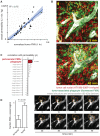
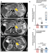
Anticancer nanotherapeutics have shown mixed results in clinical trials, raising the questions of whether imaging should be used to i) identify patients with a higher likelihood of nanoparticle accumulation, ii) assess nanotherapeutic efficacy before traditional measures show response, and iii) guide adjuvant treatments to enhance therapeutic nanoparticle (TNP) delivery. Here we review the use of a clinically approved MRI nanoparticle (ferumoxytol, FMX) to predict TNP delivery and efficacy. It is becoming increasingly apparent that nanoparticles used for imaging, despite clearly distinct physicochemical properties, often co-localize with TNP in tumors. This evidence offers the possibility of using FMX as a generic "companion diagnostic" nanoparticle for multiple TNP formulations, thus potentially allowing many of the complex regulatory and cost challenges of other approaches to be avoided.
Keywords: dextran-coated iron oxide nanoparticle; enhanced permeability and retention effect; magnetic resonance imaging; nanomedicine; personalized medicine; tumor associated macrophage..
Conflict of interest statement
Competing Interests: The authors have declared that no competing interest exists.
Figures


Similar articles
-
Predicting therapeutic nanomedicine efficacy using a companion magnetic resonance imaging nanoparticle.Sci Transl Med. 2015 Nov 18;7(314):314ra183. doi: 10.1126/scitranslmed.aac6522. Sci Transl Med. 2015. PMID: 26582898 Free PMC article.
-
Radiation therapy primes tumors for nanotherapeutic delivery via macrophage-mediated vascular bursts.Sci Transl Med. 2017 May 31;9(392):eaal0225. doi: 10.1126/scitranslmed.aal0225. Sci Transl Med. 2017. PMID: 28566423 Free PMC article.
-
Improving nanotherapy delivery and action through image-guided systems pharmacology.Theranostics. 2020 Jan 1;10(3):968-997. doi: 10.7150/thno.37215. eCollection 2020. Theranostics. 2020. PMID: 31938046 Free PMC article. Review.
-
Intraoperative assessment and postsurgical treatment of prostate cancer tumors using tumor-targeted nanoprobes.Nanotheranostics. 2021 Jan 1;5(1):57-72. doi: 10.7150/ntno.50095. eCollection 2021. Nanotheranostics. 2021. PMID: 33391975 Free PMC article.
-
Imaging-assisted anticancer nanotherapy.Theranostics. 2020 Jan 1;10(3):956-967. doi: 10.7150/thno.38288. eCollection 2020. Theranostics. 2020. PMID: 31938045 Free PMC article. Review.
Cited by
-
FGF2 engineered SPIONs attenuate tumor stroma and potentiate the effect of chemotherapy in 3D heterospheroidal model of pancreatic tumor.Nanotheranostics. 2020 Jan 1;4(1):26-39. doi: 10.7150/ntno.38092. eCollection 2020. Nanotheranostics. 2020. PMID: 31911892 Free PMC article.
-
Membrane-core nanoparticles for cancer nanomedicine.Adv Drug Deliv Rev. 2020;156:23-39. doi: 10.1016/j.addr.2020.05.005. Epub 2020 May 22. Adv Drug Deliv Rev. 2020. PMID: 32450105 Free PMC article. Review.
-
Stimuli-Responsive Iron Oxide Nanotheranostics: A Versatile and Powerful Approach for Cancer Therapy.Adv Healthc Mater. 2021 Mar;10(5):e2001044. doi: 10.1002/adhm.202001044. Epub 2020 Nov 23. Adv Healthc Mater. 2021. PMID: 33225633 Free PMC article. Review.
-
Smart cancer nanomedicine.Nat Nanotechnol. 2019 Nov;14(11):1007-1017. doi: 10.1038/s41565-019-0567-y. Epub 2019 Nov 6. Nat Nanotechnol. 2019. PMID: 31695150 Free PMC article.
-
Supermagnetic Human Serum Albumin (HSA) Nanoparticles and PLGA-Based Doxorubicin Nanoformulation: A Duet for Selective Nanotherapy.Int J Mol Sci. 2022 Dec 30;24(1):627. doi: 10.3390/ijms24010627. Int J Mol Sci. 2022. PMID: 36614071 Free PMC article.
References
-
- Juliano RL, Stamp D. Pharmacokinetics of liposome-encapsulated anti-tumor drugs. Studies with vinblastine, actinomycin D, cytosine arabinoside, and daunomycin. Biochem Pharmacol. 1978;27:21–7. - PubMed
-
- Moghimi SM, Hunter AC, Murray JC. Long-circulating and target-specific nanoparticles: theory to practice. Pharmacol Rev. 2001;53:283–318. - PubMed
-
- O'Brien ME, Wigler N, Inbar M, Rosso R, Grischke E, Santoro A. et al. Reduced cardiotoxicity and comparable efficacy in a phase III trial of pegylated liposomal doxorubicin HCl (CAELYX/Doxil) versus conventional doxorubicin for first-line treatment of metastatic breast cancer. Ann Oncol. 2004;15:440–9. - PubMed
Publication types
Grants and funding
LinkOut - more resources
Full Text Sources
Other Literature Sources

