Case Reports
. 2017 Sep;3(3):229-234.
doi: 10.1159/000458414. Epub 2017 Mar 23.

Bilateral Central Retinal Artery Occlusion Associated with Bilateral Lymphoproliferative Infiltrative Optic Neuropathy

Affiliations
Case Reports

Bilateral Central Retinal Artery Occlusion Associated with Bilateral Lymphoproliferative Infiltrative Optic Neuropathy

Philip J DeSouza et al. Ocul Oncol Pathol. 2017 Sep.

Abstract

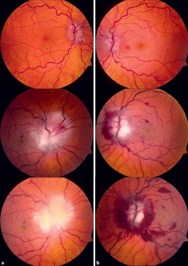
Background: Leukemic infiltration of the optic nerve is relatively rare. While previously described in acute leukemia, the infiltration in our case represents central nervous system (CNS) metastasis of Burkitt-type lymphoma that developed as a complication of solid-organ transplantation, resulting in a bilateral infiltrative optic neuropathy with sequential, bilateral central retinal artery occlusion (CRAO) and devastating vision loss.

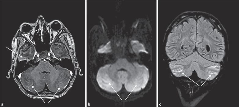
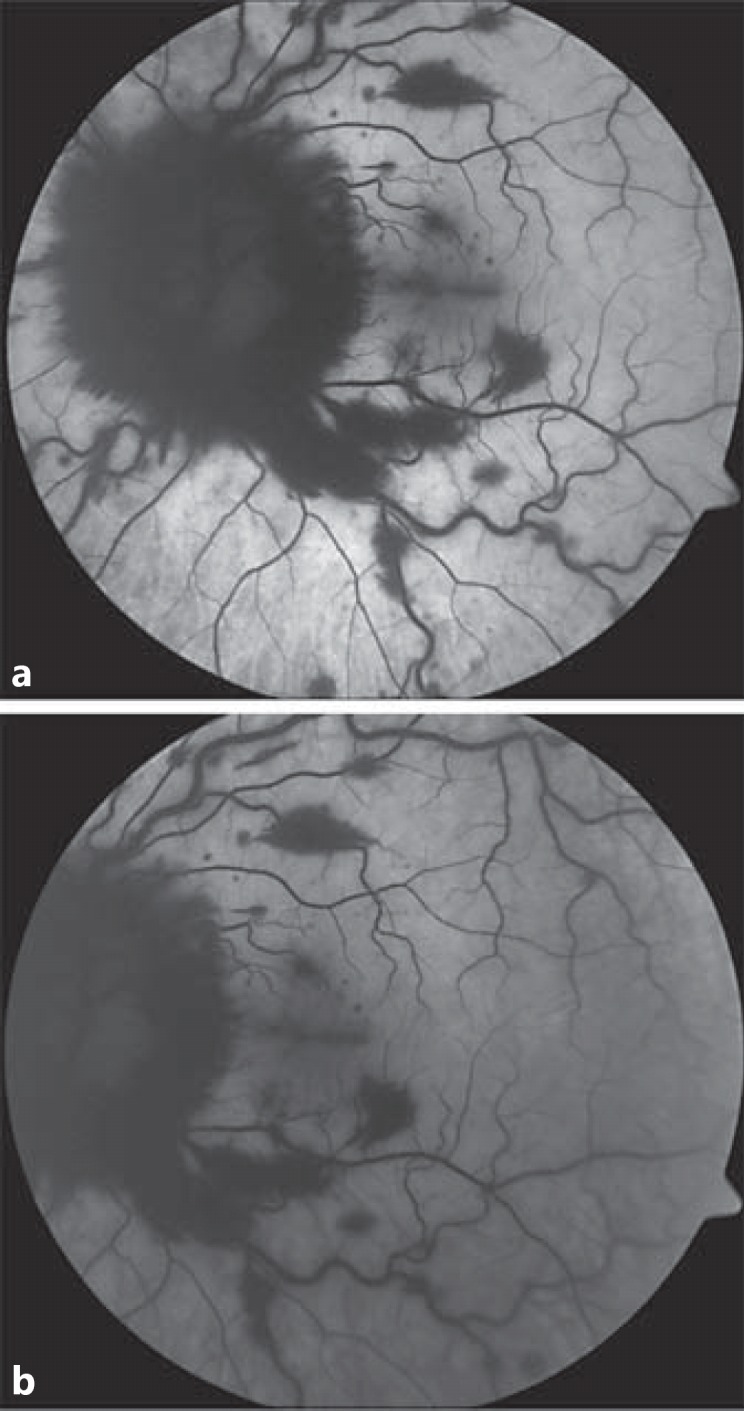
Methods: The medical record, serial ophthalmic examination findings, clinical course, and imaging including magnetic resonance imaging (MRI), fundus photographs, and fluorescein angiography of a single patient were retrospectively reviewed.

Results: MRI demonstrated multifocal cortical and leptomeningeal CNS involvement, including the left optic nerve. Serial fundus examination/photography and fluorescein angiography showed that despite urgent whole-brain irradiation and systemic chemotherapy, CNS disease progressed to bilateral optic nerve infiltration and CRAO with no light perception vision in both eyes.

Conclusion: CRAO can occur as a devastating and irreversible complication of lymphoproliferative optic nerve infiltration.

Keywords: Burkitt lymphoma; Case report; Central retinal artery occlusion; Leukemic central nervous system infiltration; Optic nerve infiltration; Post-transplant lymphoproliferative disorder.


Figures

Fig. 1
Fig. 1
Right (a) and left (b) fundus photograph on initial evaluation show bilateral blurred disc margins and hyperemic vessels. The left optic disc and macula appear white with arterial “boxcarring” and scattered hemorrhages. Right (a, middle) and left (b, middle) fundus photographs 5 days later show a fluffy, white appearance over the optic discs, consistent with prelaminar infiltrate, along with hemorrhage, particularly on the nasal side of the left optic disc. Bilateral whitening of the macula is present, and the vessels are tortuous and dilated. Right (a, bottom) and left (b, bottom) photographs 13 days after initial evaluation show progression of the infiltrates, with notably increased hemorrhage surrounding the left optic disc and retina.
Fig. 2
Fig. 2
Coronal short T1 inversion recovery magnetic resonance image showing an increased signal in the left optic nerve, consistent with optic nerve edema.
Fig. 3
Fig. 3
a Axial T1 magnetic resonance imaging volumetric interpolated breath-hold examination showing curvilinear enhancement around the folia of the cerebellum bilaterally and along the anterior aspect of the right temporal lobe consistent with a leptomeningeal process. b Axial diffusion-weighted imaging showing a hyperintense signal in the area of enhancement in the bilateral cerebellar hemispheres, suggesting hypercellular material. c Coronal T2-weighted fluid attenuated inversion recovery imaging showing a hyperintense signal filling the cerebrospinal fluid spaces around the folia of the cerebellum, suggesting proteinaceous material. Overall, the findings suggest leptomeningeal involvement of the patient's known post-transplant lymphoproliferative disorder. The hyperintense signal in the anteroinferior temporal lobes, parafalcine parietooccipital cortex, and pineal gland is not shown in the figure.
Fig. 4
Fig. 4
Fluorescein angiogram of the left eye demonstrating early (a) and late (b) hypofluorescence in the retinal vasculature, consistent with central retinal artery occlusion.

References

    1. Savani BN, Mohty M. Clinical Guide to Transplantation in Lymphoma. Chichester: John Wiley & Sons; 2015.
    1. Taylor AL, Marcus R, Bradley JA. Post-transplant lymphoproliferative disorders (PTLD) after solid organ transplantation. Crit Rev Oncol Hematol. 2005;56:155–167. - PubMed
    1. Quinlan SC, Pfeiffer RM, Morton LM, Engels EA. Risk factors for early-onset and late-onset post-transplant lymphoproliferative disorder in kidney recipients in the United States. Am J Hematol. 2011;86:206–209. - PMC - PubMed
    1. Naik S, Tayapongsak K, Robbins K, Manavi CK, Pettenati MJ, Grier DD. Burkitt's lymphoma presenting as late-onset posttransplant lymphoproliferative disorder following kidney and pancreas transplantation: case report and review of the literature. Case Rep Oncol. 2013;6:6–14. - PMC - PubMed
    1. Loren AW, Porter DL, Stadtmauer EA, Tsai DE. Post-transplant lymphoproliferative disorder: a review. Bone Marrow Transplant. 2003;31:145–155. - PubMed

Publication types

LinkOut - more resources