. 2018 Jan 1;596(1):47-66.
doi: 10.1113/JP275033. Epub 2017 Nov 23.

Transforming growth factor-β enhances Rho-kinase activity and contraction in airway smooth muscle via the nucleotide exchange factor ARHGEF1

Affiliations

Transforming growth factor-β enhances Rho-kinase activity and contraction in airway smooth muscle via the nucleotide exchange factor ARHGEF1

Yasin Shaifta et al. J Physiol. .

Abstract

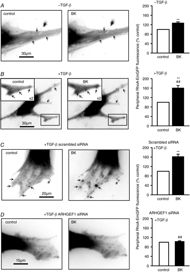
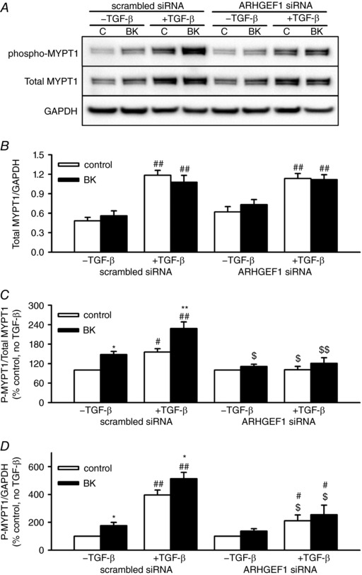
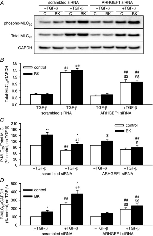
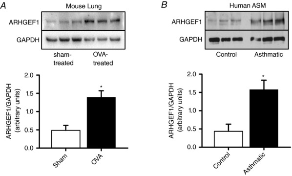
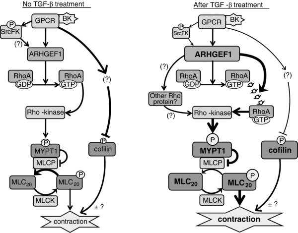
Key points: Transforming growth-factor-β (TGF-β) and RhoA/Rho-kinase are independently implicated in the airway hyper-responsiveness associated with asthma, but how these proteins interact is not fully understood. We examined the effects of pre-treatment with TGF-β on expression and activity of RhoA, Rho-kinase and ARHGEF1, an activator of RhoA, as well as on bradykinin-induced contraction, in airway smooth muscle. TGF-β enhanced bradykinin-induced RhoA translocation, Rho-kinase-dependent phosphorylation and contraction, but partially suppressed bradykinin-induced RhoA activity (RhoA-GTP content). TGF-β enhanced the expression of ARHGEF1, while a small interfering RNA against ARHGEF1 and a RhoGEF inhibitor prevented the effects of TGF-β on RhoA and Rho-kinase activity and contraction, respectively. ARHGEF1 expression was also enhanced in airway smooth muscle from asthmatic patients and ovalbumin-sensitized mice. ARHGEF1 is a key TGF-β target gene, an important regulator of Rho-kinase activity and therefore a potential therapeutic target for the treatment of asthmatic airway hyper-responsiveness.

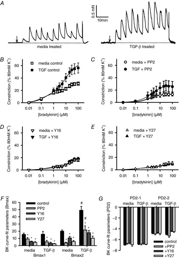
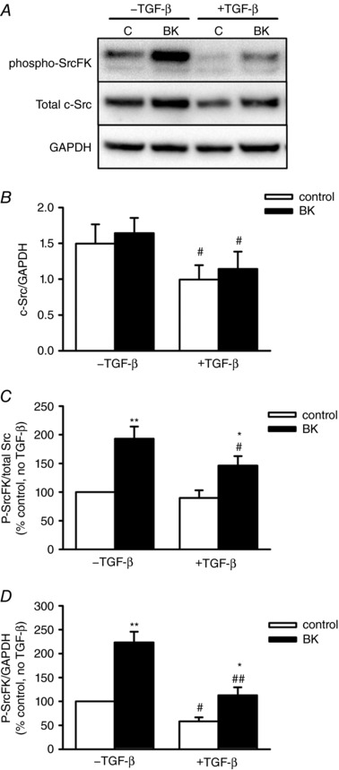
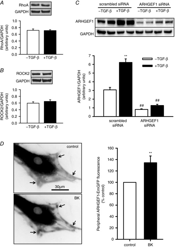
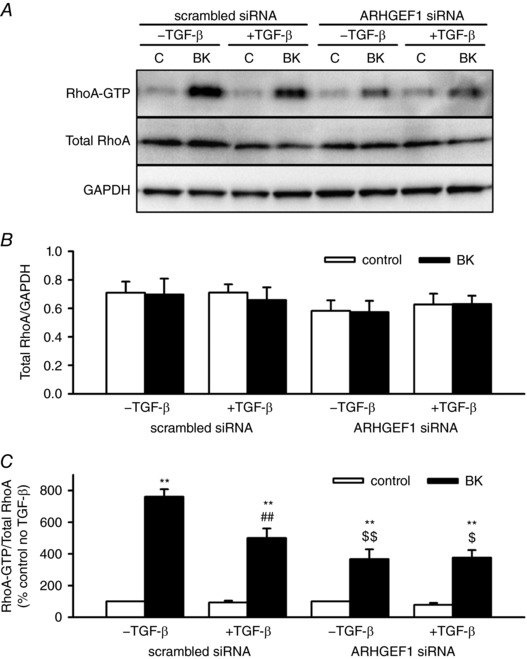
Abstract: Transforming growth factor-β (TGF-β), RhoA/Rho-kinase and Src-family kinases (SrcFK) have independently been implicated in airway hyper-responsiveness, but how they interact to regulate airway smooth muscle contractility is not fully understood. We found that TGF-β pre-treatment enhanced acute contractile responses to bradykinin (BK) in isolated rat bronchioles, and inhibitors of RhoGEFs (Y16) and Rho-kinase (Y27632), but not the SrcFK inhibitor PP2, prevented this enhancement. In cultured human airway smooth muscle cells (hASMCs), TGF-β pre-treatment enhanced the protein expression of the Rho guanine nucleotide exchange factor ARHGEF1, MLC20 , MYPT-1 and the actin-severing protein cofilin, but not of RhoA, ROCK2 or c-Src. In hASMCs, acute treatment with BK triggered subcellular translocation of ARHGEF1 and RhoA and enhanced auto-phosphorylation of SrcFK and phosphorylation of MYPT1 and MLC20 , but induced de-phosphorylation of cofilin. TGF-β pre-treatment amplified the effects of BK on RhoA translocation and MYPT1/MLC20 phosphorylation, but suppressed the effects of BK on RhoA-GTP content, SrcFK auto-phosphorylation and cofilin de-phosphorylation. In hASMCs, an ARHGEF1 small interfering RNA suppressed the effects of BK and TGF-β on RhoA-GTP content, RhoA translocation and MYPT1 and MLC20 phosphorylation, but minimally influenced the effects of TGF-β on cofilin expression and phosphorylation. ARHGEF1 expression was also enhanced in ASMCs of asthmatic patients and in lungs of ovalbumin-sensitized mice. Our data indicate that TGF-β enhances BK-induced contraction, RhoA translocation and Rho-kinase activity in airway smooth muscle largely via ARHGEF1, but independently of SrcFK and total RhoA-GTP content. A role for smooth muscle ARHGEF1 in asthmatic airway hyper-responsiveness is worthy of further investigation.

Keywords: Rho-kinase; TGF-β; airway smooth muscle.


Figures

Figure 1
Figure 1. Effects of TGF‐β and SrcFK/RhoGEF/Rho‐kinase inhibition on BK‐induced contraction
Measurement of isometric tension in isolated rat bronchioles mounted on the wire myograph. A, representative traces showing bradykinin (BK) applied cumulatively (0.01–100 μm) at 5 min intervals, after 18 h pre‐incubation in media only or in media with 30 ng ml−1 TGF‐β. Arrows indicate when the first dose of BK was applied. BE, mean contraction amplitude after TGF‐β or media pre‐incubation measured at the plateau phase of each BK application, in the absence (B, media: n = 24, TGF‐β: n = 23) or presence of 30 μm PP2 (C, media: n = 17, TGF‐β: n = 15), 30 μm Y16 (D, media: n = 9, TGF‐β: n = 9) or 10 μm Y27632 (E, media: n = 9, TGF‐β: n = 9). F and G, non‐linear regression data, showing effects of TGF‐β and inhibitors on Bmax (F) and PD2 values (G). * P < 0.05 vs. control, # P < 0.05 vs. media, two‐way ANOVA.
Figure 2
Figure 2. Effects of BK and TGF‐β on SrcFK expression and auto‐phosphorylation in hASMC
A, representative blots showing effect of BK (1 μm, 30 s) with or without prior exposure to TGF‐β (10 ng ml−1, 24 h) on phospho‐SrcFK content, total c‐Src content and GAPDH as a loading control. B, data expressed as c‐Src/GAPDH show partial inhibition of c‐Src protein expression after TGF‐β pre‐incubation (# P < 0.05 vs. –TGF‐β, n = 7). C and D, SrcFK phosphorylation is enhanced by BK (* P < 0.05, ** P < 0.01 vs. control, n = 7), but suppressed by TGF‐β (# P < 0.01, ## P < 0.01 vs. –TGF‐β, n = 7). All comparisons by two‐way ANOVA.
Figure 3
Figure 3. Effects of TGF‐β and BK on ARHGEF1 expression and translocation
AC, effects of TGF‐β incubation (10 ng ml−1, 24 h) on protein expression of RhoA (A, n = 6), Rho‐kinase (B, ROCK2, n = 5) and ARHGEF1 (C, after transfection with scrambled or ARHGEF1 siRNA, n = 9) in hASMCs, relative to GAPDH. ** P < 0.01 vs. –TGF‐β, ## P < 0.01 vs. scrambled siRNA, two‐way ANOVA. D, fluorescence imaging of live hASMCs transfected with ARHGEF1‐EmGFP. Arrows indicate peripheral regions in which ARHGEF1‐EmGFP concentrates when stimulated by addition of BK (1 μm). Data show relative changes in fluorescence in peripheral regions. ** P < 0.01 vs. control, n = 11, paired t test.
Figure 4
Figure 4. Effects of TGF‐β and ARHGEF1 siRNA on RhoA‐GTP content in hASMCs
A, representative blots showing effects of acute BK treatment (1 μm, 30 s) with or without TGF‐β pre‐treatment (10 ng ml−1, 24 h) in hASMCs transfected with either ARHGEF1 siRNA or a scrambled siRNA control on RhoA‐GTP content, total RhoA or GAPDH as a loading control. B, data expressed as total/GAPDH show no overall effect of BK, TGF‐β or ARHGEF1 siRNA on total RhoA expression. C, data expressed as RhoA‐GTP/total show significant enhancement by BK (** P < 0.01 vs. control) but partial suppression by TGF‐β (## P < 0.01, vs. –TGF‐β) and ARHGEF1 siRNA ($ P < 0.05, $$ P < 0.01 vs. scrambled siRNA). n = 7 for all data. All comparisons by three‐way ANOVA.
Figure 5
Figure 5. Effects of TGF‐β and ARHGEF1 siRNA on BK‐induced RhoA‐EmGFP translocation
Fluorescence imaging of live hASMCs transfected with RhoA‐EmGFP. A, in the absence of TGF‐β pre‐incubation, addition of BK (1 μm) enhances peripheral fluorescence, as indicated by arrows. ** P < 0.001 vs. control, n = 16. B, after TGF‐β pre‐incubation (10 ng ml−1, 24 h), response to BK is enhanced and is concentrated into distinct peripheral spots/patches, as indicated by arrows. ** P < 0.001 vs. control, ## P < 0.001 vs. –TGF‐β, n = 15. C, in hASMCs pre‐treated with TGF‐β, doubly transfected with RhoA‐EmGFP and scrambled siRNA, response to BK is similar to in B, as indicated by arrows. ** P < 0.001 vs. control, n = 28. D, in hASMCs pre‐treated with TGF‐β, co‐transfected with RhoA‐EmGFP and ARHGEF1 siRNA, BK does not trigger translocation. ## P < 0.001 vs. scrambled siRNA, n = 29. All comparisons by two‐way ANOVA.
Figure 6
Figure 6. Effects of TGF‐β and ARHGEF1 siRNA on expression and BK‐induced phosphorylation of MYPT‐1 in hASMCs
A, representative blots showing effects of acute BK treatment (1 μm, 30 s) with or without TGF‐β pre‐treatment (10 ng ml−1, 24 h) in hASMCs transfected with either ARHGEF1 siRNA, or a scrambled siRNA control, on phospho‐MYPT‐1 (Thr‐696), total MYPT1 or GAPDH as a loading control. B, data expressed as total/GAPDH show enhancement of MYPT1 protein expression by TGF‐β pre‐treatment, but no effect of ARHGEF1 siRNA or BK, ## P < 0.01 vs. –TGF‐β. Data expressed as phospho/total (C) or phospho/GAPDH (D) show significant enhancement by both BK (* P < 0.05, ** P < 0.01 vs. control) and TGF‐β (# P < 0.05, ## P < 0.01 vs. –TGF‐β) and suppression by ARHGEF1 siRNA ($ P < 0.05, $$ P < 0.01 vs. scrambled siRNA). n = 9 for all data. All comparisons by three‐way ANOVA.
Figure 7
Figure 7. Effects of TGF‐β and ARHGEF1 siRNA on expression and BK‐induced phosphorylation of MLC20 in hASMCs
A, representative blots showing effects of acute BK treatment (1 μm, 30 s) with or without TGF‐β pre‐treatment (10 ng ml−1, 24 h) in hASMCs transfected with either ARHGEF1 siRNA, or a scrambled siRNA control, on phospho‐MLC20 (Ser19), total MLC20 or GAPDH as a loading control. B, data expressed as total/GAPDH show enhancement of MLC20 protein expression by TGF‐β pre‐treatment (## P < 0.01 vs. –TGF‐β), and partial suppression of this response by ARHGEF1 siRNA ($$ P < 0.01 vs. scrambled siRNA). Data expressed as phospho/total (C) or phospho/GAPDH (D) show significant enhancement by both BK (* P < 0.05, ** P < 0.01 vs. control), effects of TGF‐β pre‐treatment (# P < 0.05, ## P < 0.01 vs. –TGF‐β) and suppression by ARHGEF1 siRNA ($ P < 0.05, $$ P < 0.01 vs. scrambled siRNA). n = 9 for all data. All comparisons by three‐way ANOVA.
Figure 8
Figure 8. Effects of BK, TGF‐β and ARHGEF1 siRNA on cofilin expression and phosphorylation in hASMCs
A, representative blots showing effects of acute BK treatment (1 μm, 30 s) with or without TGF‐β pre‐treatment (10 ng ml−1, 24 h) in hASMCs transfected with either ARHGEF1 siRNA, or a scrambled siRNA control, on phospho‐cofilin (Ser3), total cofilin or GAPDH as a loading control. B, data expressed as total/GAPDH show enhancement of cofilin protein expression by TGF‐β pre‐treatment (## P < 0.01 vs. –TGF‐β), and partial suppression of this response by ARHGEF1 siRNA ($$ P < 0.01 vs. scrambled siRNA). Data expressed as phospho/total (C) or phospho/GAPDH (D) show significant de‐phosphorylation induced by BK (** P < 0.01 vs. control), prevention of de‐phosphorylation/enhancement of basal phosphorylation by TGF‐β pre‐treatment (# P < 0.05, ## P < 0.01 vs. –TGF‐β) and partial suppression of the effects of TGF‐β by ARHGEF1 siRNA ($$ P < 0.01 vs. scrambled siRNA). n = 7 for all data. All comparisons by three‐way ANOVA.
Figure 9
Figure 9. Effects of asthma and OVA‐sensitization on lung ARHGEF‐1 protein expression
Protein expression of ARHGEF‐1 relative to GAPDH. Expression is enhanced by OVA‐sensitization in mouse lung (A, * P < 0.05 vs. sham‐treated, n = 3 in each group) and is greater in ASM of asthmatic subjects vs. healthy controls (B, * P < 0.05, n = 3 subjects in each group). All comparisons by unpaired t test.
Figure 10
Figure 10. Role of ARHGEF1, SrcFK, RhoA, Rho‐kinase and cofilin in BK‐induced contraction in ASM and the influence of TGF‐β on their relative contributions
Without TGF‐β treatment. It is established that G‐protein coupled receptor (GPCR)‐induced ASM contraction is dependent on sequential activation of RhoA and Rho‐kinase, resulting in MYPT1 phosphorylation, MLCP inhibition and enhanced MLC20 phosphorylation. We show that BK also induces SrcFK activation and translocation of ARHGEF1. RhoA translocation and activation, MYPT1 and MLC20 phosphorylation are all partially ARHGEF1‐dependent. Also, contraction is partially SrcFK‐, RhoGEF‐ and Rho‐kinase‐dependent. SrcFK possibly act upstream of ARHGEF1 (but not tested in this study). BK inhibits cofilin phosphorylation, but how this influences contraction remains to be determined. After TGF‐β treatment. Expression and BK‐induced activity of c‐Src are suppressed, while expression of ARHGEF1, MYPT1, MLC20 and cofilin are enhanced. BK‐induced RhoA activity is partially suppressed, while RhoA translocation, MYPT‐1 phosphorylation, MLC20 phosphorylation and contraction are all enhanced. Enhancement of RhoA translocation and MYPT1/MLC20 phosphorylation are partially ARHGEF1‐dependent, while enhanced contraction is Rho‐kinase and RhoGEF‐dependent. Enhanced Rho‐kinase activity may result from enhanced site‐specific RhoA translocation, to counter the reduced total RhoA activity, or from activation of another Rho‐protein (both untested in this study). Cofilin phosphorylation is enhanced, but is largely independent of ARHGEF1 and whether this contributes to enhanced contraction remains to be determined. Key: size of text/box reflects degree of protein expression. Size of ‘P’ reflects degree of phosphorylation. Thickness of lines/arrows reflects strength of effect. The row of block arrows represents translocation. (?) or (±?) denotes ‘not tested in this study’.

Similar articles

Cited by

References

    1. Albinsson S, Nordstrom I & Hellstrand P (2004). Stretch of the vascular wall induces smooth muscle differentiation by promoting actin polymerization. J Biol Chem 279, 34849–34855. - PubMed
    1. Andrianantoandro E & Pollard TD (2006). Mechanism of actin filament turnover by severing and nucleation at different concentrations of ADF/cofilin. Mol Cell 24, 13–23. - PubMed
    1. Arber S, Barbayannis FA, Hanser H, Schneider C, Stanyon CA, Bernard O & Caroni P (1998). Regulation of actin dynamics through phosphorylation of cofilin by LIM‐kinase. Nature 393, 805–809. - PubMed
    1. Atfi A, Drobetsky E, Boissonneault M, Chapdelaine A & Chevalier S (1994). Transforming growth factor β down‐regulates Src family protein tyrosine kinase signaling pathways. J Biol Chem 269, 30688–30693. - PubMed
    1. Birmingham A, Anderson E, Sullivan K, Reynolds A, Boese Q, Leake D, Karpilow J & Khvorova A (2007). A protocol for designing siRNAs with high functionality and specificity. Nat Protoc 2, 2068–2078. - PubMed

Publication types

MeSH terms

Substances